Inverted Mole Meaning: What It Is, Warning Signs, and When to See a Doctor
The term “inverted mole” is not a standard medical diagnosis, yet it is one of the more common phrases people use when they notice a mole that looks sunken, indented, or concave — or when they come across clinical language about a specific type of melanocytic lesion called an inverted type-A nevus. Understanding which meaning applies to your situation matters, because the two are very different in terms of cause, appearance, and clinical significance. This article explains both meanings clearly, describes the warning signs that warrant prompt medical attention, and outlines what a dermatologist can do to evaluate any mole that concerns you.
Key Takeaways
- “Inverted mole” has two distinct meanings: a mole that appears sunken or concave to the naked eye, and a specific histological variant called an inverted type-A nevus (also known as a clonal nevus or deep penetrating nevus).
- A mole that has changed from raised to flat or indented is a clinically significant change that warrants prompt dermatology assessment, as this pattern can be associated with basal cell carcinoma or, less commonly, melanoma.
- The inverted type-A nevus is generally considered a benign melanocytic lesion, but it can mimic melanoma under the microscope, making expert histopathological assessment essential.
- Applying the ABCDE rule — Asymmetry, Border, Color, Diameter, Evolving — and the ugly duckling sign are practical first steps in monitoring any mole at home.
- Dermoscopy, skin biopsy, and mole mapping are the main clinical tools used to evaluate and monitor moles that appear unusual or are changing.
What Does “Inverted Mole” Actually Mean?
The phrase “inverted mole” is used in two quite different contexts, and confusing them can lead to unnecessary anxiety or, conversely, a false sense of reassurance. Separating these meanings is the most important first step.
Meaning 1: A Sunken or Concave Mole (Lay Usage)
In everyday language, people use “inverted mole” to describe a mole that appears to dip inward rather than sit flush with or above the surrounding skin. A sunken or concave mole may feel like a small depression or dimple in the skin. This is the opposite of a dome-shaped or raised mole. The appearance can range from a subtle flattening to a clearly indented crater-like surface. Crucially, when a mole that was previously raised becomes flat or indented over time, this change in shape is a clinically meaningful sign that should be evaluated by a dermatologist without delay.
Meaning 2: Inverted Type-A Nevus (Clinical Usage)
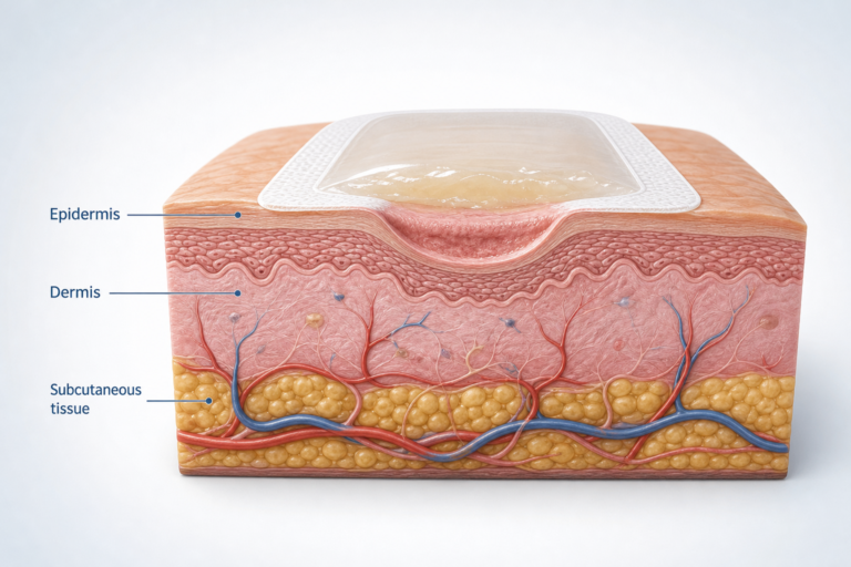
In dermatopathology — the branch of medicine that examines skin tissue under a microscope — an inverted type-A nevus (also called a clonal nevus) refers to a specific architectural pattern seen in a skin biopsy specimen. In this variant, nests of melanocytes (the pigment-producing cells of the skin) are arranged in a wedge-shaped or inverted triangle configuration, extending from the surface of the skin downward into the deeper layers of the dermis. This pattern is distinct from the typical distribution of melanocytes in a standard compound nevus or intradermal nevus. The inverted type-A nevus is closely related to the deep penetrating nevus (DPN), another benign melanocytic lesion that shares the characteristic of extending deeply into the reticular dermis or even the subcutis.
What makes the inverted type-A nevus clinically important is not that it is dangerous — it is generally classified as a benign melanocytic lesion — but that its histological features, including architectural atypia and deep melanocyte extension, can closely mimic melanoma under the microscope. This means that accurate diagnosis depends heavily on expert pathological review, and that a biopsy result describing this pattern should be discussed thoroughly with a specialist.
What Does a Sunken or Inverted Mole Look Like?
Describing the appearance of a mole that appears inverted or sunken in plain language is useful because most people are trying to assess what they can see with the naked eye, not what a pathologist sees under a microscope.
A mole that appears sunken or concave may have one or more of the following characteristics:
- A depressed or indented center, giving the mole a crater-like or dimpled appearance
- Edges that appear raised relative to the center, creating a rim around the depression
- A surface that feels rough, pebbly, or uneven rather than smooth
- Color that may be tan, brown, dark brown, or blue-black, sometimes with variation across the lesion
- Borders that may be irregular, scalloped, or poorly defined
- A size that may be larger than a typical benign mole (greater than 6 mm in diameter)
It is important to note that a sunken center is not exclusive to moles. Basal cell carcinoma (BCC), the most common form of skin cancer, classically presents as a pearly or translucent lesion with a depressed or ulcerated center and rolled edges. BCC is frequently mistaken for a mole, particularly in its early stages. If a lesion that you thought was a mole develops a sunken center, bleeds easily, or fails to heal, basal cell carcinoma should be considered and a dermatologist consulted promptly.
When a Mole Changes: The Significance of a Mole That Has Become Sunken
A mole that has changed from raised to flat or indented is one of the more concerning patterns in dermatology. Stable moles — those that have looked the same for years — are generally reassuring. A mole evolving or changing in shape, surface texture, or elevation is a different matter entirely.
Several processes can cause a previously raised mole to become sunken or flattened:
- Regression: Some melanomas and atypical moles undergo partial regression, where the immune system attacks part of the lesion. This can cause areas of the mole to flatten, develop a white or grey discolouration, or appear sunken. Regression within a pigmented lesion is considered a warning sign rather than a reassuring one.
- Basal cell carcinoma: As noted above, BCC can develop within or adjacent to a pre-existing lesion and may cause a sunken, ulcerated, or crusted appearance.
- Trauma or inflammation: Physical trauma, repeated friction, or inflammation can temporarily alter the surface of a mole. However, any change that persists beyond a few weeks should be assessed professionally.
- Spontaneous involution: Occasionally, a benign mole may flatten naturally over time as part of the normal lifecycle of a nevus. This is more common in older adults and is generally gradual and symmetrical.
The key clinical principle is this: a mole that has changed — in any direction — warrants professional evaluation. The change itself is the warning sign, regardless of whether the mole looks obviously abnormal to the untrained eye.
Applying the ABCDE Rule to an Inverted or Changing Mole
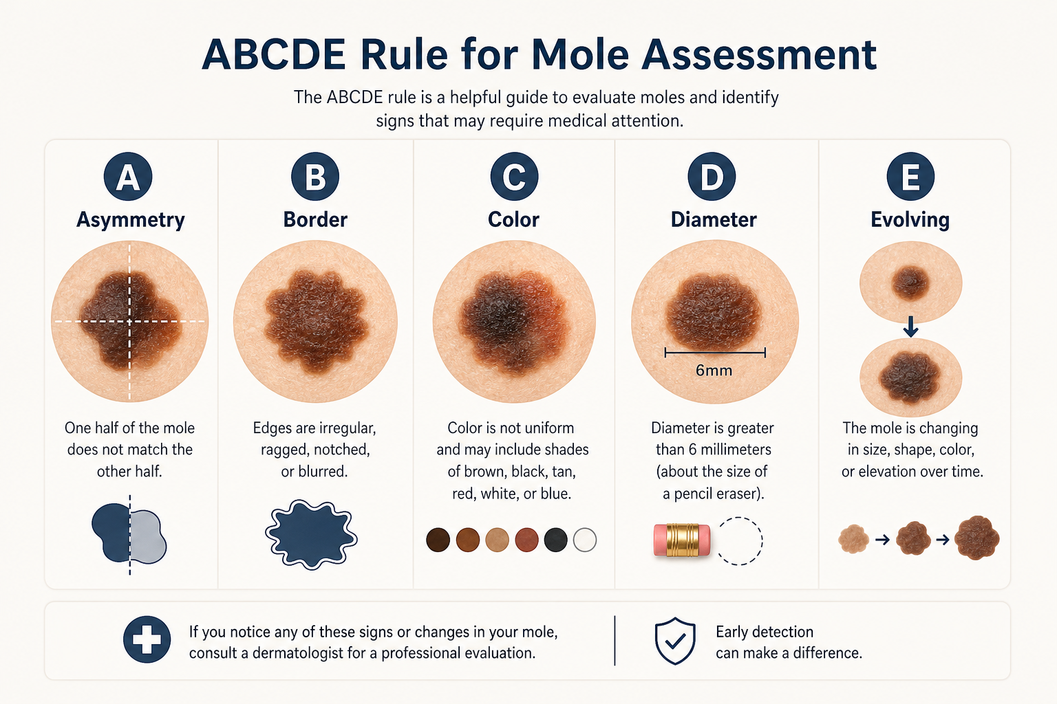
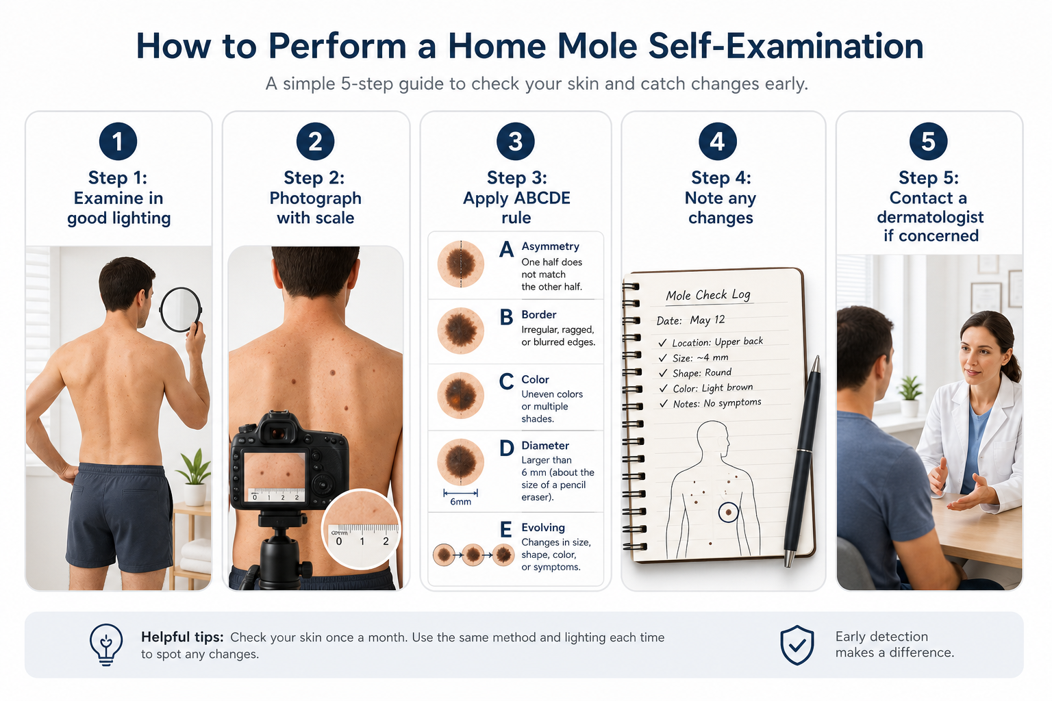
The ABCDE rule is a widely used framework for identifying moles that may require further investigation. It was developed to help both patients and clinicians identify features associated with melanoma and other concerning skin lesions. Applying it to a mole that appears sunken or has recently changed is a practical starting point.
- A — Asymmetry: One half of the mole does not match the other. A sunken center that affects only part of the mole creates asymmetry.
- B — Border: The edges are irregular, ragged, notched, or blurred. A mole with a raised rim and sunken center may have an irregular border.
- C — Color: Multiple shades of brown, black, red, white, or blue within the same lesion. Color variation within a sunken mole is a significant warning sign.
- D — Diameter: A mole larger than 6 mm (approximately the size of a pencil eraser) is more likely to warrant assessment, though melanomas can be smaller.
- E — Evolving: Any change in size, shape, color, elevation, or surface — including a mole becoming sunken — is the most important criterion. A mole evolving or changing should always be assessed by a dermatologist.
Alongside the ABCDE rule, the ugly duckling sign is a useful complementary approach. This means looking for a mole that simply looks different from all the others on your body — the one that stands out. An inverted or sunken mole that looks unlike your other moles is worth having checked, even if it does not tick every ABCDE box.
Inverted Type-A Nevus and Deep Penetrating Nevus: A Plain-Language Explanation
For those who have received a biopsy result mentioning an inverted type-A nevus, a clonal nevus, or a deep penetrating nevus (DPN), understanding what these terms mean in practical terms is important.
A deep penetrating nevus is a benign melanocytic lesion that tends to appear as a darkly pigmented, sometimes blue-black papule or nodule, most commonly on the face, upper trunk, or proximal limbs. It is typically well-defined and may be present from childhood or early adulthood. The defining histological feature is the extension of melanocytes in a wedge-shaped or inverted triangle pattern deep into the reticular dermis, sometimes reaching the subcutis. This deep extension, combined with features such as architectural atypia and pigmented spindle cells, is what causes the DPN to resemble melanoma under the microscope.
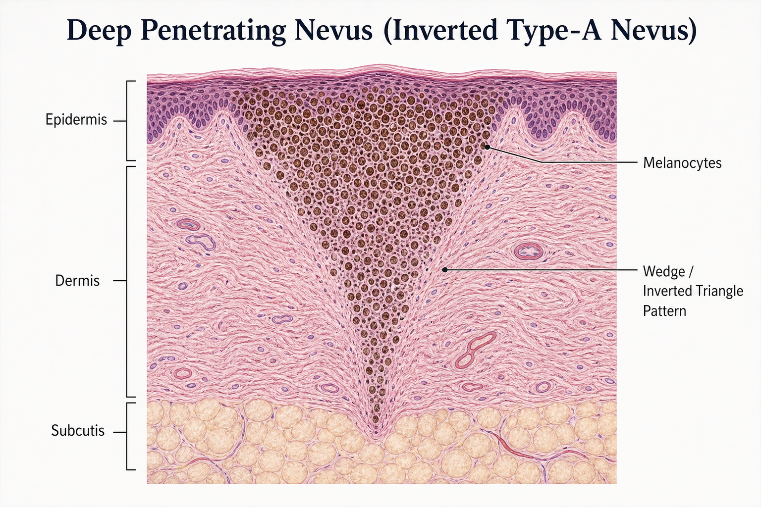
The inverted type-A nevus is a closely related entity. Research published in dermatopathology literature, including studies referenced by the Mayo Clinic and available through PubMed, describes the inverted type-A nevus as a variant in which large nests of type-A melanocytes (pale, epithelioid cells) are arranged in the characteristic inverted triangle or wedge configuration. Despite the alarming histological appearance, the clinical behaviour of both the DPN and the inverted type-A nevus is generally benign, with low rates of recurrence after excision and no established association with malignant transformation in the majority of cases.
However, because these lesions can genuinely mimic melanoma — and because distinguishing them from melanoma requires expert histopathological assessment — a biopsy result describing either entity should be reviewed by an experienced dermatopathologist. In some cases, a second opinion or additional immunohistochemical staining may be recommended before a final management decision is made.
Inverted Mole vs. Normal Mole vs. Melanoma: Key Differences
| Feature | Typical Benign Mole | Sunken / Inverted Mole | Melanoma |
|---|---|---|---|
| Shape | Round or oval, symmetrical | May be asymmetrical; sunken center | Asymmetrical, irregular outline |
| Border | Smooth, well-defined | May be raised rim with irregular edge | Ragged, notched, or blurred |
| Color | Uniform tan or brown | May show color variation | Multiple shades; may include red, white, or blue-black |
| Surface | Smooth or slightly raised | Depressed, concave, or crater-like | May be ulcerated, bleeding, or crusted |
| Change over time | Stable for years | May represent a recent change | Actively evolving |
| Clinical urgency | Routine monitoring | Prompt dermatology assessment advised | Urgent dermatology referral required |
How Is an Inverted or Unusual Mole Diagnosed?
When you visit a dermatologist with a mole that appears sunken, has changed, or is otherwise concerning, the assessment typically involves several steps.
Clinical Examination
The dermatologist will first examine the mole visually, assessing its size, shape, color, surface texture, and borders. They will also ask about any changes you have noticed, how long the mole has been present, and whether you have a personal or family history of melanoma or atypical mole syndrome (also called dysplastic nevus syndrome).
Dermoscopy
Dermoscopy — the use of a handheld dermatoscope that illuminates and magnifies the skin — allows the clinician to examine structures within the mole that are not visible to the naked eye. Dermoscopy significantly improves the accuracy of clinical diagnosis and can help distinguish between a benign melanocytic lesion, a dysplastic nevus, and a melanoma. It is a standard part of mole assessment in most dermatology settings.
Skin Biopsy
If dermoscopy or clinical examination raises concern, a skin biopsy will be recommended. This involves removing all or part of the mole under local anaesthetic and sending the tissue to a pathology laboratory for histopathological analysis. The biopsy report will describe the architectural and cellular features of the lesion, including whether architectural atypia or cytologic atypia is present, and will classify the lesion as benign, mildly atypical, moderately atypical, severely atypical, or malignant. For lesions such as the inverted type-A nevus or deep penetrating nevus, expert pathological review is particularly important given the potential for misclassification.
Mole Mapping
For patients with a high mole count, a personal or family history of melanoma, or atypical mole syndrome, mole mapping — a systematic photographic record of all moles on the body — may be recommended. Mole mapping allows clinicians to detect subtle changes over time that might not be apparent during a single examination. Follow-up skin examinations are typically scheduled every six to twelve months for higher-risk patients.
Risk Factors That Increase Concern
Not every person with a sunken or unusual mole faces the same level of risk. Several factors increase the likelihood that a changing mole warrants more urgent attention:
- Fair skin, light eyes, or light hair, which are associated with greater UV sensitivity
- A history of significant sun exposure or sunburn, particularly in childhood
- A personal or family history of melanoma or other skin cancers
- A high total mole count (generally considered to be 50 or more moles)
- A diagnosis of atypical mole syndrome or dysplastic nevus syndrome
- New moles appearing after the age of 40, or any mole changing rapidly at any age
- Immunosuppression due to medication or medical conditions
The presence of one or more of these risk factors does not mean that a changing mole is malignant, but it does mean that professional evaluation should not be delayed.
Can an Inverted Mole Be Removed?
Whether a mole should be removed depends on the clinical assessment rather than on appearance alone. Removal is generally recommended when a biopsy confirms melanoma, when a lesion shows severe atypia, or when a lesion cannot be reliably distinguished from melanoma on clinical and dermoscopic grounds. For lesions confirmed as benign — including the inverted type-A nevus and deep penetrating nevus — surgical excision may still be recommended to obtain clear margins and to prevent diagnostic uncertainty in the future, particularly if the lesion is in a location that makes monitoring difficult.
For moles that appear sunken but are confirmed as benign on biopsy, watchful waiting with regular follow-up skin examinations may be appropriate. The dermatologist will advise on the most suitable management plan based on the individual’s risk profile, the specific histological findings, and the location and size of the lesion.
Cosmetic removal of moles — for example, by laser or shave excision — is generally not recommended for moles that have not been properly assessed, as it removes the tissue needed for histopathological diagnosis and can make future monitoring more difficult.
How to Monitor a Mole at Home
Regular self-examination is a useful complement to professional skin checks, not a replacement for them. The following steps are particularly relevant for monitoring a mole that appears to be changing or that has a sunken or unusual appearance:
- Examine your skin in good lighting, using a full-length mirror and a hand mirror for hard-to-see areas such as the back, scalp, and behind the ears.
- Photograph any mole that concerns you, including a reference object such as a ruler or coin for scale. Repeat the photograph at the same angle and lighting conditions at monthly intervals.
- Apply the ABCDE rule to each mole you are monitoring, paying particular attention to the E (Evolving) criterion — any change in shape, surface, or elevation.
- Note whether the mole that was previously raised has become flat or sunken, whether it has developed a new color, or whether it has started to itch, bleed, or crust.
- If you notice any of these changes, do not wait for your next scheduled appointment — contact a dermatologist promptly.
Frequently Asked Questions
Is a sunken mole always a sign of cancer?
No. A sunken or concave mole is not automatically a sign of cancer. However, a mole that has changed from raised to flat or indented is a clinically significant change that should be assessed by a dermatologist. The cause may be benign — such as natural involution of a nevus — but basal cell carcinoma, melanoma regression, and other concerning processes can also produce this appearance. Professional evaluation is the only way to determine the cause with confidence.
What is the difference between an inverted type-A nevus and melanoma?
An inverted type-A nevus is a benign melanocytic lesion, while melanoma is a malignant skin cancer. The difficulty is that the inverted type-A nevus can closely mimic melanoma under the microscope due to its deep extension of melanocytes and architectural atypia. Distinguishing between the two requires expert histopathological assessment, and in some cases additional immunohistochemical staining. Clinically, the inverted type-A nevus does not behave like melanoma and does not carry the same prognosis.
Should I be worried if my mole changed shape?
Any mole that is evolving or changing — in shape, color, size, surface texture, or elevation — should be assessed by a dermatologist. Change is the most important warning sign in mole monitoring. This does not mean that every changing mole is malignant, but it does mean that professional evaluation should not be postponed.
What does it mean if a raised mole becomes flat or sunken?
A mole that changes from raised to flat or sunken may be undergoing natural involution, but it may also indicate regression within a melanoma, the development of basal cell carcinoma, or another process that warrants investigation. This specific change — a previously raised mole becoming concave or indented — is one of the patterns that dermatologists consider a prompt referral indicator. Do not assume the change is harmless without a professional assessment.
What is a deep penetrating nevus and is it dangerous?
A deep penetrating nevus (DPN) is a benign melanocytic lesion characterized by deeply extending melanocytes arranged in a wedge-shaped pattern within the dermis. It is generally considered non-malignant, but because it can resemble melanoma histologically, accurate diagnosis by an experienced dermatopathologist is essential. Management typically involves surgical excision to confirm the diagnosis and obtain clear margins.
How often should I have my moles checked?
For most adults, an annual full-body skin examination by a dermatologist is a reasonable baseline. Patients with a high mole count, a personal or family history of melanoma, atypical mole syndrome, or other risk factors may be advised to attend every six months and to undergo mole mapping. Between professional checks, monthly self-examination is a useful habit.
Can a mole be removed without a biopsy?
Cosmetic removal of a mole without prior histopathological assessment is generally not recommended for any mole that has changed, appears unusual, or has features that concern you or your clinician. Removing a mole without sending the tissue for analysis means that a potential melanoma or severely atypical lesion could be missed. Any mole removed for medical reasons should routinely be sent for pathological examination.
What to Do If You Are Concerned About an Inverted or Changing Mole
If you have noticed a mole that appears sunken, concave, or indented — or a mole that has changed in any way — the most important step is to arrange a professional skin examination with a dermatologist rather than attempting to self-diagnose. The term “inverted mole” covers a range of appearances and clinical entities, from the entirely benign inverted type-A nevus to changes that may indicate basal cell carcinoma or melanoma. No visual description, online guide, or self-examination tool can replace a clinical assessment that includes dermoscopy and, where indicated, a skin biopsy.
Early detection remains one of the most effective factors in improving outcomes for skin cancer. A mole that turns out to be benign after assessment provides genuine reassurance; a mole that is caught early when it is malignant is far more treatable than one identified at a later stage. If a mole is worrying you, acting promptly is always the right decision. A qualified dermatologist can provide an accurate diagnosis, explain your individual risk profile, and recommend the most appropriate management — whether that is watchful waiting, mole mapping, or surgical excision.