Interpreting Mole Biopsy Results: A Plain-English Guide to Your Pathology Report
Receiving mole biopsy results can feel overwhelming, particularly when the pathology report is written in clinical language that was never designed for patients. This guide translates the most important terms — from benign melanocytic naevus to Breslow thickness — into plain English, explains what each result category means for your health, and outlines the next steps your dermatologist is likely to discuss with you.
Key Takeaways
- Most mole biopsies return a benign result, meaning no further treatment is required beyond routine skin checks.
- An atypical or dysplastic nevus is not melanoma, but the grade of atypia (mild, moderate, or severe) influences whether re-excision is recommended.
- Clear margins mean the mole was completely removed; positive margins mean cells were present at the cut edge and further assessment may be needed.
- For melanoma diagnoses, Breslow thickness is the single most important prognostic measurement and directly determines surgical margin recommendations.
- If your result is unexpected or sits on a borderline between categories, requesting a second pathology opinion is a reasonable and accepted step.
What Is a Pathology Report and Who Writes It?
When a mole is removed — whether by shave biopsy, punch biopsy, or excisional biopsy — the tissue is sent to a laboratory where it is processed, thinly sliced, stained, and examined under a microscope. The clinician who performs this examination is a dermatopathologist: a doctor with specialist training in both dermatology and pathology. Their written findings form the pathology report, which is then sent to your dermatologist or treating clinician.
The report is a formal document, not a letter written for patients. It uses standardised terminology so that any clinician reading it can understand the findings precisely. This is why the language can seem dense or alarming at first glance. Understanding the structure of the report — and the meaning of its key fields — makes it considerably easier to have an informed conversation with your doctor.
Turnaround time for mole biopsy results is typically five to ten working days, though this can vary. Results may take longer when the dermatopathologist requests special stains, immunohistochemistry (additional laboratory tests to identify specific cell proteins), or an internal second opinion within the laboratory. If your result is taking longer than expected, this does not automatically indicate a serious finding — it often reflects the thoroughness of the review process.
How Biopsy Type Affects Your Report
The technique used to remove your mole influences what the pathology report can and cannot tell you. An excisional biopsy removes the entire lesion with a small surrounding margin of normal skin, giving the dermatopathologist the most complete picture of the mole’s architecture and the clearest assessment of margin status. A punch biopsy removes a cylindrical core of tissue and is useful for sampling but may not capture the full lesion. A shave biopsy slices the mole horizontally at skin level and is efficient for superficial lesions, but it can transect the base of a deeper mole.
This last point matters for Breslow thickness measurement. If a shave biopsy cuts through the bottom of a lesion, the report may state that the Breslow depth is “at least” a certain number of millimetres — meaning the true depth could be greater than what was measured. This qualifier is clinically significant and is one reason your dermatologist may recommend a wider excision or a different biopsy approach if the initial result raises concern.
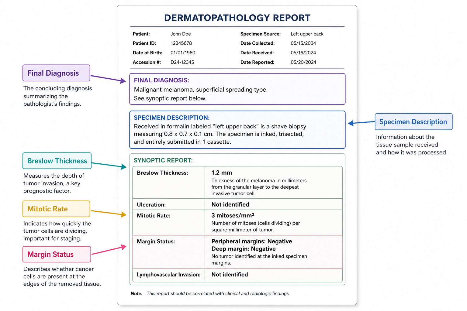
Reading Your Pathology Report: Key Sections Explained
Most pathology reports follow a consistent structure. Knowing where to look helps you understand the findings before your follow-up appointment.
Final Diagnosis
This is the most important section and usually appears near the top of the report. It states the dermatopathologist’s conclusion in one or two lines — for example, “benign melanocytic naevus” or “dysplastic nevus with moderate atypia, margins clear.” Start here when reading your report.
Specimen Description and Anatomic Site
This section records where on the body the mole was taken from, the size of the tissue sample, and a brief description of its appearance. It confirms the laboratory received the correct specimen.
Margin Status
The margin status field records whether the mole’s cells extend to the edge of the removed tissue. Clear margins (also called negative margins) mean a rim of normal tissue surrounds the mole — it has been completely excised. Positive margins mean mole cells were found at the cut edge, indicating that some tissue may remain in the skin.
Breslow Thickness
This measurement, recorded in millimetres, describes how deeply a melanocytic lesion extends into the skin. It is the dominant prognostic factor for melanoma and directly determines the recommended surgical margin for any further surgery.
Mitotic Rate
The mitotic rate (sometimes called the mitotic index) counts the number of actively dividing cells per square millimetre of tissue. A higher mitotic rate in a melanoma indicates more rapid cell division and is associated with a less favourable prognosis. This field is most relevant when a melanoma diagnosis has been made.
Ulceration
The report will note whether ulceration — breakdown of the skin surface overlying the tumour — is present or absent. Ulceration is an independent prognostic factor in melanoma and influences AJCC staging.
The Three Main Result Categories
| Category | What It Means | Typical Next Step |
|---|---|---|
| Benign | Normal mole cells; no abnormal features | Routine skin surveillance; no further treatment needed |
| Atypical / Dysplastic | Abnormal cell features present; graded mild, moderate, or severe | Depends on grade and margin status; may require re-excision |
| Malignant | Melanoma (in situ or invasive) confirmed | Wide local excision; staging; possible sentinel lymph node biopsy |
Benign Melanocytic Naevus
A result of benign melanocytic naevus means the mole is composed of normal melanocytes (the pigment-producing cells of the skin) arranged in a typical pattern with no features of concern. This is the most common mole biopsy result. No further treatment is required, though your dermatologist will likely recommend ongoing skin surveillance — particularly if you have many moles or a personal or family history of skin cancer.
Atypical Mole and Dysplastic Nevus: The Grey Area
An atypical mole or dysplastic nevus is not melanoma. It is a mole with abnormal features that fall between a clearly benign nevus and a malignant lesion. The dermatopathologist grades the degree of abnormality as mild, moderate, or severe atypia, based on two types of cellular change: architectural atypia (abnormal arrangement of cells within the mole) and cytologic atypia (abnormal appearance of individual cells).
The grade of atypia, combined with the margin status, determines what happens next. This is a clinical nuance that many patient-facing resources do not explain clearly:
- Mild atypia with clear margins: Observation is generally appropriate. Re-excision is not routinely recommended.
- Mild atypia with positive margins: Re-excision is often not required, as the residual tissue is unlikely to progress. Your dermatologist will advise based on the specific findings.
- Moderate atypia with positive margins: Re-excision is commonly recommended, though clinical judgement applies.
- Severe atypia with positive margins: Re-excision is typically recommended, usually with 2 mm surgical margins, because severe dysplasia carries a meaningfully higher risk of progression if residual tissue remains.
A positive margin result does not mean cancer has spread. It means some abnormal cells may remain at the biopsy site and your clinician needs to assess whether further removal is warranted.
Melanoma In Situ
Melanoma in situ means melanoma cells are confined to the outermost layer of the skin (the epidermis) and have not invaded deeper tissue. It is the earliest stage of melanoma and is highly treatable. The standard treatment is wide local excision (WLE) with surgical margins of 0.5 to 1 centimetre of surrounding normal skin. Because the cells have not penetrated beyond the epidermis, sentinel lymph node biopsy is not indicated at this stage.
Invasive Melanoma
Invasive melanoma means the tumour has grown beyond the epidermis into the dermis or deeper layers. The management of invasive melanoma is guided primarily by Breslow thickness, with additional input from mitotic rate, ulceration, and other prognostic factors recorded in the pathology report.
Understanding Breslow Thickness and Surgical Margins
Breslow thickness is measured in millimetres from the top of the epidermis to the deepest tumour cell visible under the microscope. It is the single most powerful predictor of melanoma outcome and directly determines the recommended width of the surgical margin at wide local excision.
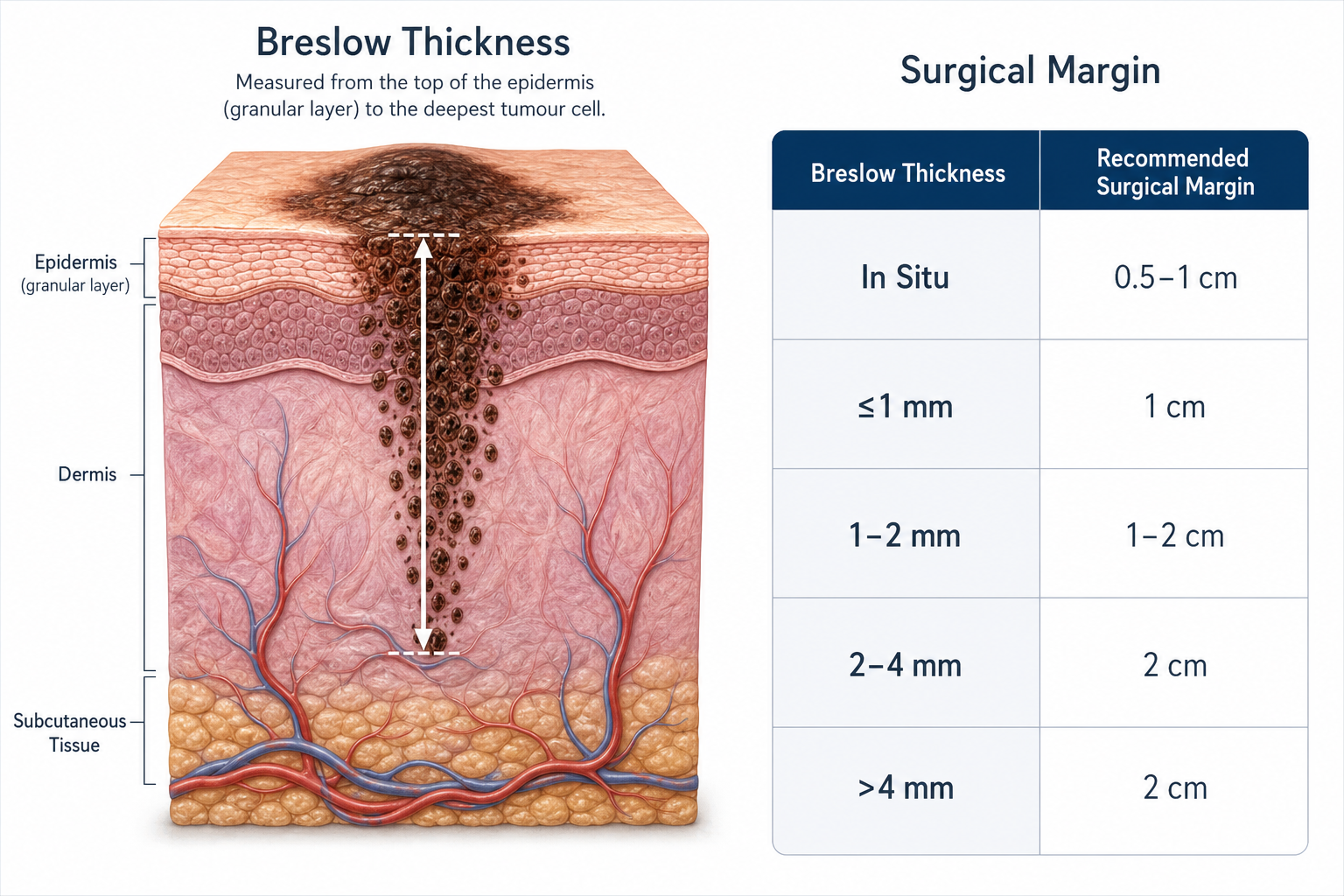
| Breslow Thickness | Recommended Surgical Margin | Sentinel Lymph Node Biopsy |
|---|---|---|
| Melanoma in situ | 0.5–1 cm | Not indicated |
| ≤1.0 mm (T1) | 1 cm | Generally not indicated; discuss with specialist |
| 1.01–2.0 mm (T2) | 1–2 cm | May be discussed depending on other features |
| 2.01–4.0 mm (T3) | 2 cm | Typically recommended |
| >4.0 mm (T4) | 2 cm | Typically recommended |
When a shave biopsy has transected the base of the lesion, the report may record Breslow thickness as “at least” a given measurement. This qualifier signals that the true depth is unknown and that a wider excision — which will provide a complete specimen — is needed to determine the actual thickness and guide definitive management.
Clark Level, AJCC Staging, and Other Report Fields
Older pathology reports and some current laboratories still record Clark level, a five-point scale describing the anatomical depth of melanoma invasion (from the epidermis at Level I to the subcutaneous fat at Level V). Clark level was historically used as a prognostic factor, but the AJCC 8th edition staging system — the current international standard — relies primarily on Breslow thickness, ulceration, and mitotic rate rather than Clark level. If Clark level appears on your report, your dermatologist can explain its relevance in the context of your specific findings.
AJCC staging (also written as TNM or pTNM staging) classifies melanoma using three components: pT (primary tumour depth and features), pN (lymph node involvement), and pM (distant metastasis). The staging notation on your report — for example, pT1a or pT2b — encodes Breslow thickness, ulceration status, and mitotic rate into a single classification that guides treatment planning and follow-up intensity. Your oncology or dermatology team will explain your specific stage and what it means for your care pathway.
Other fields that may appear in a melanoma report include lymphovascular invasion (whether tumour cells have entered blood or lymph vessels near the primary site), regression (areas where the immune system has partially destroyed tumour cells), and tumour-infiltrating lymphocytes (immune cells within the tumour, which may carry a favourable prognostic signal). These features contribute to the overall clinical picture but are interpreted alongside Breslow thickness rather than in isolation.
Sentinel Lymph Node Biopsy: When Is It Discussed?
Sentinel lymph node biopsy (SLNB) is a surgical procedure that samples the first lymph node(s) draining the area of the primary melanoma. It is used to determine whether melanoma cells have spread to the regional lymphatic system — information that influences staging and treatment decisions. SLNB is generally discussed for melanomas with a Breslow thickness greater than 1 mm, and sometimes for thinner melanomas with high-risk features such as ulceration or a high mitotic rate. A positive sentinel node result typically leads to a discussion about adjuvant (additional) therapy and further lymph node assessment.
Non-Melanoma Findings in a Mole Biopsy
Although most mole biopsies are sent to assess a pigmented lesion for melanocytic abnormality, the pathology report occasionally identifies a different type of skin cancer entirely. Basal cell carcinoma (BCC) and squamous cell carcinoma (SCC) can arise within or adjacent to a pigmented lesion and may be found incidentally. If your report mentions BCC or SCC rather than a melanocytic diagnosis, your dermatologist will explain the specific management pathway, which differs from that for melanoma or dysplastic nevi.
Recurrent Nevus: A Specific Situation Worth Knowing
If a mole was previously removed incompletely and has regrown at the same site, the new biopsy may show features that look alarming under the microscope — a phenomenon known as a recurrent nevus or pseudomelanoma. The regrowth pattern can mimic melanoma histologically, which is why it is important to inform your dermatologist (and ensure the pathology request form notes) that the site was previously biopsied. With this clinical context, the dermatopathologist can distinguish a recurrent benign nevus from a genuinely malignant lesion. Failing to provide this history can lead to an unnecessarily alarming report.
When to Consider a Second Pathology Opinion
Requesting a second opinion on a pathology report is a legitimate and accepted step in dermatological care. It is particularly worth considering in the following situations:
- The diagnosis is unexpected given the clinical appearance of the mole.
- The report describes a borderline finding — for example, severe dysplasia versus melanoma in situ — where the distinction significantly changes management.
- The report uses qualifying language such as “cannot exclude” or “features suggestive of” without a definitive conclusion.
- You are being referred for a major surgical procedure and want independent confirmation of the diagnosis.
A second opinion involves sending the original tissue slides (not just the written report) to another accredited dermatopathologist. Your dermatologist can facilitate this referral, and it does not imply distrust of the original laboratory — it is a standard quality assurance step in complex or borderline cases.
What Happens Next Based on Your Result
The pathway after receiving mole biopsy results depends on the diagnosis, the grade of any atypia, and the margin status. The following outlines the general approach, though individual management is always tailored by your treating clinician:
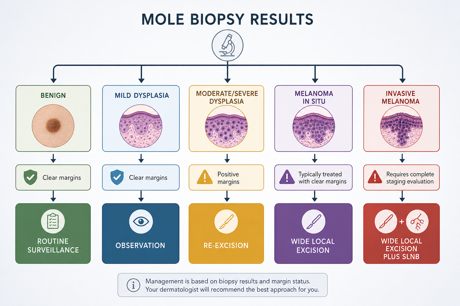
- Benign melanocytic naevus, clear margins: No further treatment. Routine skin surveillance recommended, with frequency depending on your overall risk profile.
- Mild dysplastic nevus, clear margins: Observation. No re-excision required in most cases.
- Moderate or severe dysplastic nevus, positive margins: Re-excision is commonly recommended to ensure complete removal.
- Melanoma in situ: Wide local excision with 0.5–1 cm margins. Regular dermatologist follow-up and full skin surveillance.
- Invasive melanoma ≤1 mm: Wide local excision with 1 cm margins. Sentinel lymph node biopsy discussed on a case-by-case basis.
- Invasive melanoma >1 mm: Wide local excision with margins determined by Breslow thickness. Sentinel lymph node biopsy typically recommended. Referral to a melanoma multidisciplinary team.
- Positive sentinel lymph node: Discussion of adjuvant systemic therapy and further lymph node assessment with an oncologist.
Questions to Ask Your Dermatologist
Bringing a prepared list of questions to your follow-up appointment helps ensure you leave with a clear understanding of your result and the recommended plan. The following questions are relevant for most mole biopsy consultations:
- What is the exact diagnosis, and what does it mean for my health?
- Were the margins clear, and does that change what treatment I need?
- If re-excision is recommended, how wide will the margins be and why?
- If melanoma was found, what is the Breslow thickness and what stage does that correspond to?
- Do I need a sentinel lymph node biopsy, and what would a positive result mean?
- How often should I have skin checks going forward?
- Should I inform family members to have their skin checked?
- Is a second pathology opinion appropriate in my case?
Frequently Asked Questions
How long does it take to get mole biopsy results?
Most mole biopsy results are available within five to ten working days. Results can take longer when the laboratory requests additional staining techniques, immunohistochemistry, or an internal second review. A longer wait does not necessarily indicate a more serious finding.
Does a positive margin mean the cancer has spread?
No. A positive margin means mole cells were present at the edge of the removed tissue — it describes the local completeness of removal, not whether cells have travelled to other parts of the body. Whether re-excision is needed depends on the type and grade of cells found at the margin.
My report says “moderate atypia.” Do I definitely need re-excision?
Not necessarily. The recommendation depends on whether the margins are clear or positive. Moderate atypia with clear margins may be managed with observation in some clinical contexts. Moderate atypia with positive margins more commonly leads to a recommendation for re-excision. Your dermatologist will advise based on the full report and your individual circumstances.
What does “clear margins” mean — does it confirm the mole is completely gone?
Clear margins (also called negative margins) mean that a rim of normal tissue surrounds the removed mole in the specimen examined. This is the best indicator that the lesion has been completely excised. However, the adequacy of clear margins also depends on how wide that rim is, which is why the recommended margin width varies by diagnosis.
Is a dysplastic nevus the same as skin cancer?
No. A dysplastic nevus is an atypical mole with abnormal features, but it is not melanoma or any other form of skin cancer. People with multiple dysplastic nevi do have a statistically higher lifetime risk of developing melanoma, which is why regular skin surveillance is recommended — but the presence of a dysplastic nevus does not mean cancer is present or inevitable.
What does the Clark level on my report mean?
Clark level is a five-point scale (I through V) describing how deeply a melanoma has invaded the layers of the skin. It still appears on many pathology reports but has largely been replaced by Breslow thickness as the primary prognostic measurement in current AJCC staging guidelines. If Clark level appears on your report, your dermatologist can explain whether it is influencing your management plan.
Can I request a second opinion on my pathology report?
Yes, and it is a reasonable step — particularly for borderline, unexpected, or complex diagnoses. A second opinion requires sending the original tissue slides to another accredited dermatopathologist. Your treating clinician can arrange this referral.
Making Sense of Your Mole Biopsy Report: The Practical Takeaway
A mole biopsy pathology report contains a significant amount of clinical information compressed into a short document. The most important fields to locate are the final diagnosis, the margin status, and — if melanoma is present — the Breslow thickness. These three elements together determine the recommended next steps in the vast majority of cases.
For most people, mole biopsy results will confirm a benign melanocytic naevus requiring no further treatment. For those with an atypical or dysplastic nevus, the grade of atypia and the margin status together guide whether observation or re-excision is appropriate — and the distinction between mild, moderate, and severe atypia matters considerably. For the smaller number of patients whose results indicate melanoma, the Breslow thickness drives surgical planning, sentinel lymph node biopsy decisions, and AJCC staging.
If your result is unclear, unexpected, or sits on a diagnostic borderline, a second pathology opinion is a legitimate and accepted option. Whatever your result, a follow-up appointment with a dermatologist who can review the full report in the context of your clinical history remains the most important next step. Skin cancer biopsy interpretation is a specialist skill, and the written report is the starting point for a conversation — not a substitute for one.