Cryotherapy for Moles: How Freezing Works, Who It Suits, and What to Expect
Cryotherapy for moles — sometimes called cryosurgery — uses extreme cold to destroy the cells that make up a mole. It is a minimally invasive outpatient procedure that appeals to patients who want to avoid a surgical incision, but it comes with important limitations that are not always clearly explained. This guide covers how the treatment works, which moles may be suitable, what the procedure involves, realistic risks including incomplete removal, and how cryotherapy compares to surgical excision and other alternatives.
Key Takeaways
- Cryotherapy destroys mole tissue using liquid nitrogen or nitrous oxide, but it does not produce a tissue sample, meaning a histopathology examination to rule out skin cancer is not possible after treatment.
- The procedure works best on flat, superficial benign moles; raised or deeper moles have a significantly higher rate of incomplete removal and recurrence.
- Pigmentation changes — particularly hypopigmentation — are common, especially in people with darker skin tones, and may be permanent.
- Surgical excision remains the clinical gold standard for mole removal because it allows pathology testing and achieves more reliable clearance.
- Any mole that has not been assessed by a qualified clinician using dermoscopy should not be treated with cryotherapy until a benign diagnosis is confirmed.
What Is Cryotherapy for Moles?
Cryotherapy — also referred to as cryosurgery in clinical settings — is a technique that applies a cryogen, most commonly liquid nitrogen at approximately −196°C, directly to a skin lesion to freeze and destroy its cells. In aesthetic and minor surgery clinics, handheld devices such as the CryoPen use nitrous oxide as the cryogen, reaching approximately −89°C. Both approaches work by inducing a controlled frostbite effect within the target tissue, but the lower temperature of liquid nitrogen allows a deeper freeze, which matters when treating anything other than a very superficial skin lesion.
Moles — medically known as melanocytic nevi — are clusters of pigment-producing cells called melanocytes. A benign melanocytic nevus poses no health risk, but patients may seek cosmetic mole removal for aesthetic reasons or because a mole catches on clothing or causes mild irritation. Cryotherapy is one of several methods used for this purpose, alongside surgical excision, shave excision, laser removal, and electrocautery.
How Does Cryotherapy Work on Mole Tissue?
When liquid nitrogen or nitrous oxide is applied to a mole, the extreme cold causes rapid ice crystal formation inside the cells. These ice crystals rupture cell membranes, leading to cellular destruction. The freeze-thaw cycle — the process of freezing the tissue and then allowing it to thaw — is the key mechanism. A second freeze-thaw cycle is often applied immediately after the first to increase the depth and completeness of cell death. The treated tissue subsequently blisters, scabs, and gradually sloughs away over one to six weeks as the body clears the damaged cells.
The depth of tissue destruction depends on the cryogen used, the duration of freezing (typically five to fifteen seconds per cycle), the application method — whether a cryoprobe, cotton swab, or precision spray tip — and the number of freeze-thaw cycles applied. Liquid nitrogen mole removal can reach deeper dermal layers than nitrous oxide devices, which is relevant when considering whether a particular mole is suitable for cryotherapy at all.
Which Moles May Be Suitable for Cryotherapy?
Not all moles respond equally well to freezing, and patient selection is the most important factor in determining whether cryotherapy is an appropriate choice. The procedure is most likely to achieve satisfactory clearance on flat, superficial moles — technically described as junctional nevi, where the melanocyte clusters sit at the junction between the epidermis and dermis. These lesions are shallower, and the freeze can reach all of the relevant cells.
Raised moles — compound nevi or intradermal nevi — extend deeper into the dermis. Because the freeze may not penetrate to the full depth of the lesion, the risk of incomplete removal is substantially higher. Published data suggest that incomplete removal rates for moles treated with cryotherapy range from approximately 30 to 50 percent, compared with much lower rates for surgical excision. Incomplete removal means that some melanocytes survive, which can lead to mole regrowth — sometimes with an irregular appearance that may be difficult to reassess clinically.
Before any cryotherapy treatment, a qualified clinician should perform a dermoscopy assessment — an examination using a handheld dermatoscope that magnifies the mole and reveals structural features invisible to the naked eye. Dermoscopy helps identify features that may suggest a mole is not straightforwardly benign and should be sent for histopathology rather than destroyed. Any mole showing features that raise concern under dermoscopy should not be treated with cryotherapy.
The ABCDE Warning Signs
Patients should be aware of the ABCDE warning signs before seeking any form of mole removal. These criteria — Asymmetry, Border irregularity, Colour variation, Diameter greater than 6 mm, and Evolution or change over time — are used by clinicians to identify moles that require urgent assessment rather than cosmetic treatment. A mole that has recently changed in size, shape, or colour, bleeds without trauma, or itches persistently should be evaluated by a doctor before any removal procedure is considered. Cryotherapy is appropriate only for confirmed benign lesions.
The Critical Problem: No Tissue Sample After Cryotherapy
One of the most important limitations of cryotherapy for moles is that the procedure destroys the tissue entirely. Unlike surgical excision, which removes the mole intact and allows it to be sent to a laboratory as a pathology specimen, cryotherapy leaves nothing to examine. This means that if the mole contained early melanoma or atypical cells, that diagnosis would be missed entirely — and the patient would have no way of knowing.
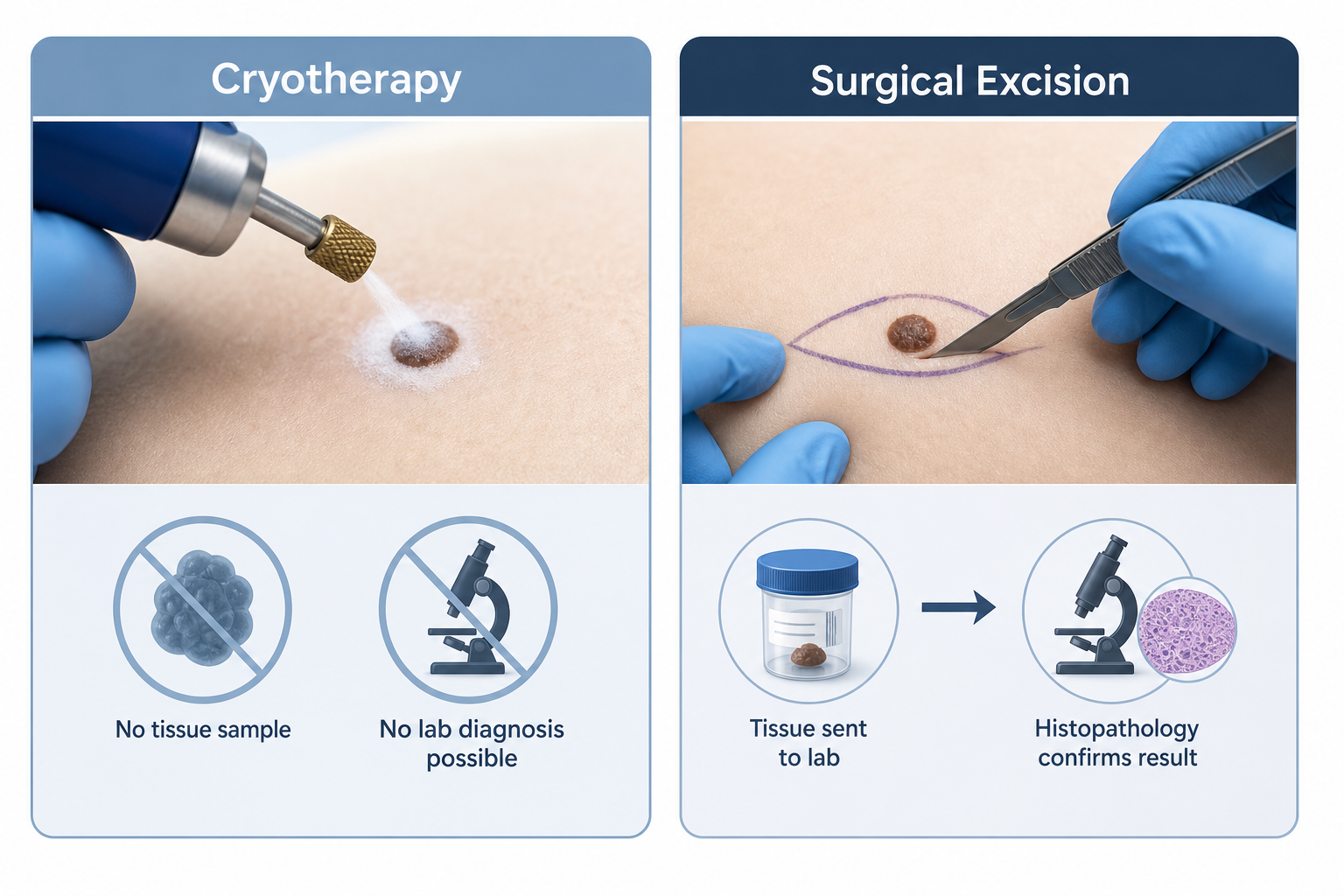
This is not a theoretical concern. Melanoma, the most serious form of skin cancer, can occasionally resemble a benign mole, particularly in its early stages. Even experienced clinicians using dermoscopy cannot achieve 100 percent diagnostic certainty in every case. Surgical excision followed by histopathology examination remains the only way to confirm that a removed mole was entirely benign. Patients choosing cryotherapy are accepting that this confirmation will not be available, which is why a thorough pre-treatment dermoscopy assessment by a qualified practitioner is essential rather than optional.
If a mole treated with cryotherapy later turns out to have been cancerous — for example, if a similar lesion elsewhere is biopsied and found to be melanoma — the destroyed tissue cannot be retrieved or re-examined. This can delay diagnosis and complicate treatment planning. For this reason, many dermatologists and minor surgery specialists recommend surgical excision as the preferred approach for any mole where there is even a small degree of diagnostic uncertainty.
What Happens During a Cryotherapy Appointment?
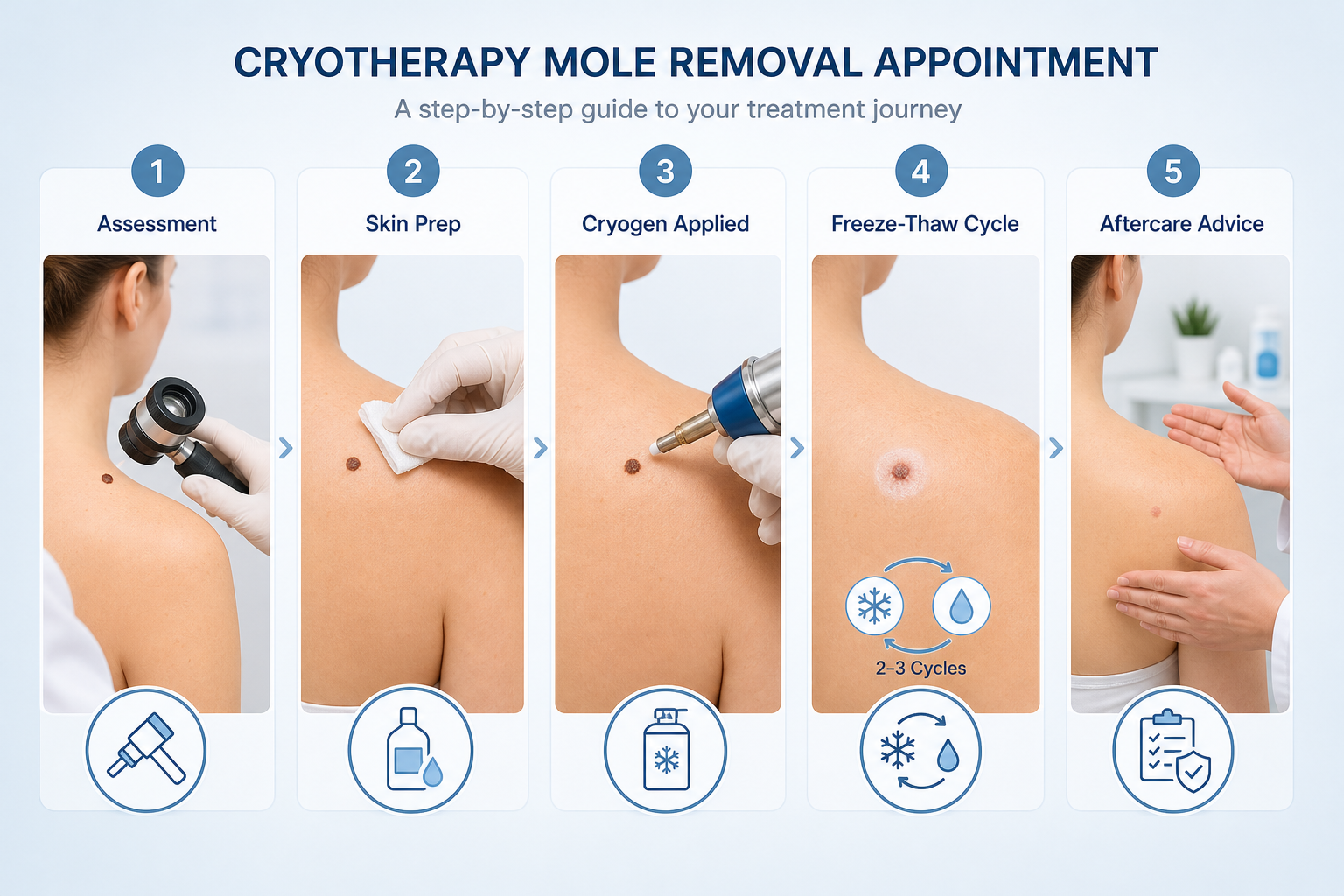
Cryotherapy for mole removal is performed as an outpatient procedure, typically in a dermatology clinic, minor surgery centre, or aesthetic practice. The appointment usually takes between fifteen and thirty minutes, depending on the number of lesions being treated and the method used.
- Assessment and consent: The clinician examines the mole, often using a dermatoscope, and confirms it is suitable for cryotherapy. The risks, limitations, and alternatives are discussed, and the patient provides informed consent.
- Skin preparation: The area is cleaned. Local anaesthetic is not usually required for small superficial lesions, though some practitioners offer a topical numbing cream for patient comfort.
- Application of the cryogen: Liquid nitrogen is applied using a spray device or cryoprobe, or a handheld nitrous oxide device such as the CryoPen is used. The clinician applies the cryogen for a controlled duration — typically five to fifteen seconds — while maintaining a precise margin around the mole.
- Freeze-thaw cycle: The tissue is allowed to thaw, then a second freeze-thaw cycle is often applied to improve the depth of cell destruction.
- Immediate aftercare advice: The clinician explains what to expect over the following days and weeks, including blistering, scabbing, and healing, and provides written aftercare instructions.
Most patients describe the sensation as a sharp stinging or burning feeling during the freeze, followed by a throbbing ache for several minutes afterwards. Discomfort is generally mild and short-lived for small lesions.
Risks and Side Effects
Cryotherapy carries a predictable set of side effects and a smaller number of less common risks. Understanding these before treatment helps patients make an informed decision.
Common Side Effects
- Blistering and scabbing: Blister formation occurs in the majority of cases — estimates suggest 60 to 80 percent of treated sites develop a blister within 24 to 48 hours. This is a normal part of the healing process. The blister should not be deliberately burst, as doing so increases infection risk.
- Redness and swelling: Localised inflammation is expected immediately after treatment and typically settles within a few days.
- Mild discomfort: Tenderness at the treated site is common for the first day or two.
Pigmentation Changes
Hypopigmentation — a lightening of the skin at the treated site — is one of the most frequently reported longer-term side effects of cryotherapy. It occurs because the melanocytes responsible for skin colour are destroyed along with the mole cells. In people with lighter skin tones, a pale patch may be subtle and fade over months. In people with darker skin tones — particularly those with Fitzpatrick skin types IV to VI — hypopigmentation can be more pronounced, more visible, and potentially permanent. Hyperpigmentation, a darkening of the surrounding skin, can also occur, particularly during the healing phase. Patients with darker skin tones should discuss these risks in detail with their clinician before proceeding.
Scarring
Cryotherapy carries a lower risk of scarring than surgical excision in many cases, but scarring is not impossible. Deeper freezes, larger lesions, and individual healing responses all influence the outcome. Some patients develop a flat, pale scar at the treated site, which may be less noticeable than a surgical scar but can still be permanent.
Incomplete Removal and Recurrence
As noted above, incomplete removal is a significant limitation of cryotherapy for moles. When some melanocytes survive the freeze, the mole can regrow — sometimes within weeks or months. Recurrent moles may look different from the original, with irregular borders or uneven pigmentation, which can make clinical reassessment more difficult. If recurrence occurs, the clinician needs to reassess the lesion carefully before deciding whether a further cryotherapy session or a different approach is more appropriate.
Less Common Risks
- Infection: Rare, occurring in fewer than approximately 2 percent of cases when aftercare instructions are followed correctly.
- Nerve damage: Uncommon, but possible when treating moles over areas where superficial nerves run close to the skin surface.
- Delayed diagnosis of skin cancer: As discussed, the absence of a pathology specimen means that a cancerous lesion could be missed.
CryoPen vs Liquid Nitrogen: Does the Device Matter?
The choice of cryogen and device affects the depth of freeze and therefore the suitability of cryotherapy for different mole types. Liquid nitrogen, used in spray or cryoprobe systems, reaches approximately −196°C and can achieve a deeper freeze than handheld nitrous oxide devices such as the CryoPen or Freezpen, which reach approximately −89°C.
For very superficial, flat lesions — including some solar lentigines, seborrheic keratoses, and junctional nevi — nitrous oxide cryotherapy may achieve adequate results. For moles with any dermal component, the shallower freeze of a nitrous oxide device may be insufficient to destroy all of the melanocyte clusters, increasing the likelihood of incomplete removal. Patients should ask their clinician which device will be used and why it is appropriate for their specific lesion, rather than assuming that all cryotherapy devices are equivalent.
Aftercare and Recovery
Following cryotherapy, the treated site goes through a predictable healing sequence. Understanding this timeline helps patients manage expectations and recognise when something may need clinical attention.
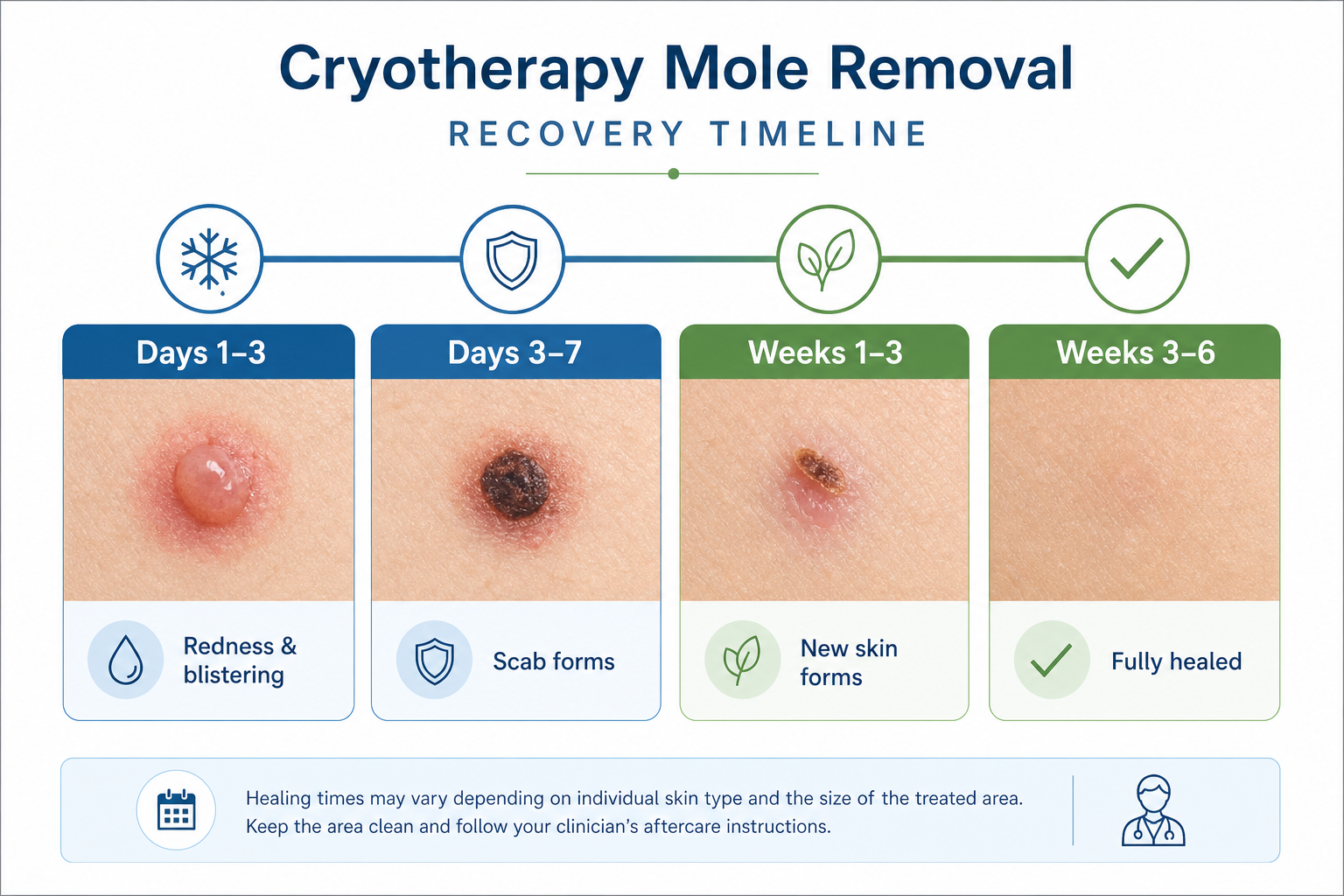
- Days 1–3: Redness, swelling, and blister formation are expected. Keep the area clean and dry. Do not burst blisters deliberately.
- Days 3–7: The blister may burst naturally or begin to dry. A scab forms over the treated area. Do not pick or remove the scab prematurely, as this increases the risk of scarring and infection.
- Weeks 1–3: The scab gradually lifts as new skin forms underneath. The area may appear pink or pale.
- Weeks 3–6: Most sites are fully healed by six weeks, though pigmentation changes may take longer to stabilise.
Specific aftercare instructions typically include: cleaning the site gently with mild soap and water once daily; applying a thin layer of petroleum jelly or a recommended wound dressing if advised; avoiding swimming, saunas, and prolonged water exposure until the site is healed; and applying a broad-spectrum SPF 30 or higher sunscreen to the healed area daily for at least three months to reduce the risk of post-inflammatory pigmentation changes. If the site becomes increasingly red, warm, swollen, or begins to discharge pus, contact the treating clinic promptly as these may be signs of infection.
A follow-up appointment is advisable at four to eight weeks to confirm that the mole has cleared completely and that the healing is progressing as expected. If there is any sign of mole regrowth at follow-up, the clinician should reassess the lesion before deciding on next steps.
When Multiple Sessions May Be Needed
A single cryotherapy session is not always sufficient to achieve complete clearance, particularly for raised or pigmented moles. If residual mole tissue is visible at follow-up, a second session may be recommended, typically after a minimum of four to eight weeks to allow the skin to heal fully. In some cases, a third session may be required. Patients should be aware of this possibility before starting treatment, as multiple sessions affect both the overall cost and the total healing time. If two sessions have not achieved clearance, switching to surgical excision — which also provides the histopathology specimen that cryotherapy cannot — is often the more appropriate course of action.
Cryotherapy vs Surgical Excision and Other Alternatives
| Method | Mechanism | Produces Pathology Specimen | Suitable Mole Types | Clearance Rate | Downtime | Scarring Risk | Approximate Cost (UK) |
|---|---|---|---|---|---|---|---|
| Cryotherapy (liquid nitrogen) | Freeze-thaw cell destruction | No | Flat, superficial benign moles | Moderate; 30–50% incomplete removal for moles | 1–6 weeks healing | Low to moderate | £95–£300 |
| Surgical excision | Scalpel removal with sutures | Yes | All mole types including raised and deep | High when adequate margins taken | 1–2 weeks; suture removal at 7–14 days | Linear scar expected | £200–£600+ |
| Shave excision | Blade shaves mole flush with skin | Yes (partial) | Raised moles; not suitable for flat pigmented lesions | Good for raised lesions; recurrence possible | 1–3 weeks | Low; flat scar | £150–£400 |
| Laser removal (CO₂ / IPL) | Light energy vaporises tissue | No | Superficial pigmented lesions; not all moles | Variable; multiple sessions often needed | 1–4 weeks | Low to moderate | £100–£400 per session |
| Electrocautery / diathermy | Electrical current destroys tissue | No | Superficial raised lesions | Moderate; recurrence possible | 1–3 weeks | Low to moderate | £100–£350 |
Surgical excision is widely regarded as the clinical gold standard for mole removal because it removes the lesion intact, allows histopathology examination of the pathology specimen, and achieves reliable clearance when adequate margins are taken. The trade-off is a linear scar and a slightly longer recovery involving suture removal. For patients who are primarily concerned about cosmetic outcome and whose mole has been confirmed as benign through dermoscopy assessment, cryotherapy or shave excision may be reasonable alternatives — but the absence of a tissue sample remains an important consideration in every case.
A Note on At-Home Freezing Kits
Over-the-counter freezing products designed for wart removal are not appropriate for use on moles. These products typically use dimethyl ether propane mixtures that reach temperatures of approximately −57°C — significantly warmer than liquid nitrogen and insufficient to destroy the deeper melanocyte clusters in a mole. More importantly, using an at-home kit on a mole bypasses any clinical assessment entirely, meaning that a potentially cancerous lesion could be treated without diagnosis. At-home freezing kits should not be used on moles under any circumstances.
NHS vs Private Treatment in the UK
In the UK, the NHS will generally fund mole removal only when there is a clinical reason — for example, when a mole is suspected of being cancerous or pre-cancerous, or when it causes significant functional problems. Cosmetic mole removal is not routinely funded by the NHS. Patients with a mole that has changed or shows ABCDE warning signs should see their GP first, who can refer them through the two-week-wait urgent skin cancer pathway if appropriate. For moles that are confirmed benign but the patient wishes to have removed for cosmetic reasons, private treatment is the usual route, with cryotherapy typically costing between £95 and £300 per lesion depending on the clinic and the method used.
Frequently Asked Questions
Does cryotherapy hurt?
Most patients experience a sharp stinging sensation during the freeze and a throbbing ache for several minutes afterwards. For small superficial lesions, local anaesthetic is not usually required, though some clinics offer topical numbing cream. Discomfort is generally manageable and short-lived.
Will cryotherapy leave a scar?
Cryotherapy carries a lower scarring risk than surgical excision in many cases, but scarring is possible, particularly with deeper freezes or larger lesions. The treated area may heal with a pale, flat patch rather than a raised scar. Individual healing responses vary, and pigmentation changes at the site may be permanent.
Can a mole grow back after cryotherapy?
Yes. Mole regrowth after cryotherapy is a recognised limitation, with incomplete removal rates estimated at 30 to 50 percent for moles. If some melanocytes survive the freeze, the mole can recur — sometimes with a different appearance. A follow-up appointment is important to check for recurrence and reassess the site if needed.
How many sessions will I need?
Some moles clear after a single session, but a second or third session may be required, particularly for raised or pigmented lesions. Sessions are typically spaced four to eight weeks apart to allow full healing between treatments. If multiple sessions do not achieve clearance, surgical excision may be recommended.
Is cryotherapy safe for darker skin tones?
Cryotherapy can be performed on darker skin tones, but the risk of hypopigmentation — a permanent lightening of the treated area — is higher and more visible in people with Fitzpatrick skin types IV to VI. This should be discussed thoroughly with the clinician before treatment, as the cosmetic outcome may not be acceptable to all patients.
Can I have cryotherapy if my mole has not been checked?
No. Any mole should be assessed by a qualified clinician using dermoscopy before cryotherapy is performed. Treating an unassessed mole risks destroying tissue that may have been cancerous, eliminating the possibility of a histopathology diagnosis and potentially delaying cancer treatment.
What is the difference between cryotherapy and at-home wart-freezing kits?
At-home wart-freezing products do not reach temperatures low enough to destroy mole tissue effectively, and they provide no clinical assessment. They are not suitable for use on moles and should not be used as an alternative to professional treatment.
Is Cryotherapy the Right Choice for Your Mole?
Cryotherapy for moles is a practical, minimally invasive option for patients with confirmed benign, superficial lesions who prefer to avoid a surgical incision. It is quick, does not usually require local anaesthetic, and carries a relatively low risk of significant scarring for suitable candidates. However, it is not the right choice for every mole or every patient.
The absence of a pathology specimen is the most significant limitation. Any mole that has not been thoroughly assessed — or where there is any diagnostic uncertainty — should be removed by surgical excision so that histopathology can confirm the diagnosis. The realistic incomplete removal rate of 30 to 50 percent for moles means that cryotherapy is less reliable than surgical excision for achieving complete clearance, and patients should be prepared for the possibility of needing more than one session or switching to a different method.
Patients with darker skin tones should weigh the risk of permanent hypopigmentation carefully, and anyone considering a handheld nitrous oxide device such as the CryoPen should confirm with their clinician that the device is appropriate for their specific lesion depth. A thorough dermoscopy assessment before treatment, clear aftercare instructions, and a scheduled follow-up appointment are the minimum standards that a responsible cryotherapy provider should offer. If you are considering cosmetic mole removal, a consultation with a qualified dermatologist or minor surgery specialist is the appropriate starting point.