Mole on Finger: Warning Signs, Types, and When to See a Dermatologist
A mole on the finger is usually a benign nevus — a harmless cluster of pigment-producing melanocytes — but the fingers and surrounding nail area are also sites where rarer, more serious skin cancers can develop. Understanding the difference between a mole that simply needs monitoring and one that warrants prompt dermatological assessment can make a meaningful difference to outcomes. This guide covers the types of moles that appear on fingers, the specific warning signs to watch for, the unique risks associated with finger and nail anatomy, and the practical steps you can take to monitor your skin safely at home.
Key Takeaways
- Most moles on fingers are benign, but the fingers and nail area are known sites for acral lentiginous melanoma, a rarer melanoma subtype that does not require sun exposure to develop.
- The ABCDE rule — Asymmetry, Border, Color, Diameter, Evolution — is the standard framework for assessing whether a mole needs professional evaluation.
- A dark streak running lengthwise under the nail (longitudinal melanonychia) is not the same as a mole on the finger surface and requires separate, specialist assessment.
- People with darker skin tones are disproportionately affected by acral lentiginous melanoma and subungual melanoma, making awareness particularly important across all skin types.
- Any mole on the finger that is new after age 30, changing, bleeding, itching persistently, or looks different from your other moles should be assessed by a dermatologist without delay.
Can You Get a Mole on Your Finger?
Yes — moles can appear anywhere on the body where melanocytes are present, and the fingers are no exception. Melanocytes are the pigment-producing cells distributed throughout the skin, including on the dorsal (back) surface of the fingers, the palmar side of the finger, the fingertips, knuckles, and the periungual area surrounding the nail. Most finger moles are acquired nevi that develop during childhood or early adulthood in response to genetic factors and, on the sun-exposed dorsal surface, cumulative UV exposure. A congenital mole — one present from birth — can also appear on the finger, though this is less common.
Finger moles are sometimes confused with other skin lesions. Common look-alikes include viral warts (verrucae), seborrheic keratoses, dermatofibromas, blood blisters caused by trauma, and contact dermatitis reactions to metals in rings or cleaning products. Because the fingers are high-contact areas exposed to friction, chemicals, and jewelry, distinguishing a true mole from an irritated or reactive skin lesion is not always straightforward without professional assessment.
Types of Moles That Can Appear on Fingers
Not all moles carry the same risk profile. Understanding the main types helps frame what a dermatologist will be looking for during an examination.
- Common acquired nevus: A flat or slightly raised, evenly pigmented mole that develops after birth. These are the most frequently seen moles on the fingers and are generally benign.
- Congenital mole (congenital nevus): Present from birth. Larger congenital nevi carry a modestly elevated lifetime risk of melanoma compared with acquired moles, though the absolute risk for small congenital nevi remains low.
- Dysplastic nevus (atypical mole): A mole with irregular borders, uneven color, or a size greater than 6 mm. A single dysplastic nevus is not automatically dangerous, but people with multiple dysplastic nevi — sometimes called dysplastic nevus syndrome — have a higher lifetime risk of melanoma and benefit from regular mole mapping and dermatological surveillance.
- Spitz nevus: A pink or reddish raised lesion that can resemble melanoma under the microscope. Spitz nevi are more common in children and young adults and typically require biopsy to confirm their nature.
- Blue nevus: A blue-grey mole caused by melanocytes located deeper in the dermis. These are usually benign but can occasionally be confused with nodular melanoma.
The ABCDE Rule for Finger Moles
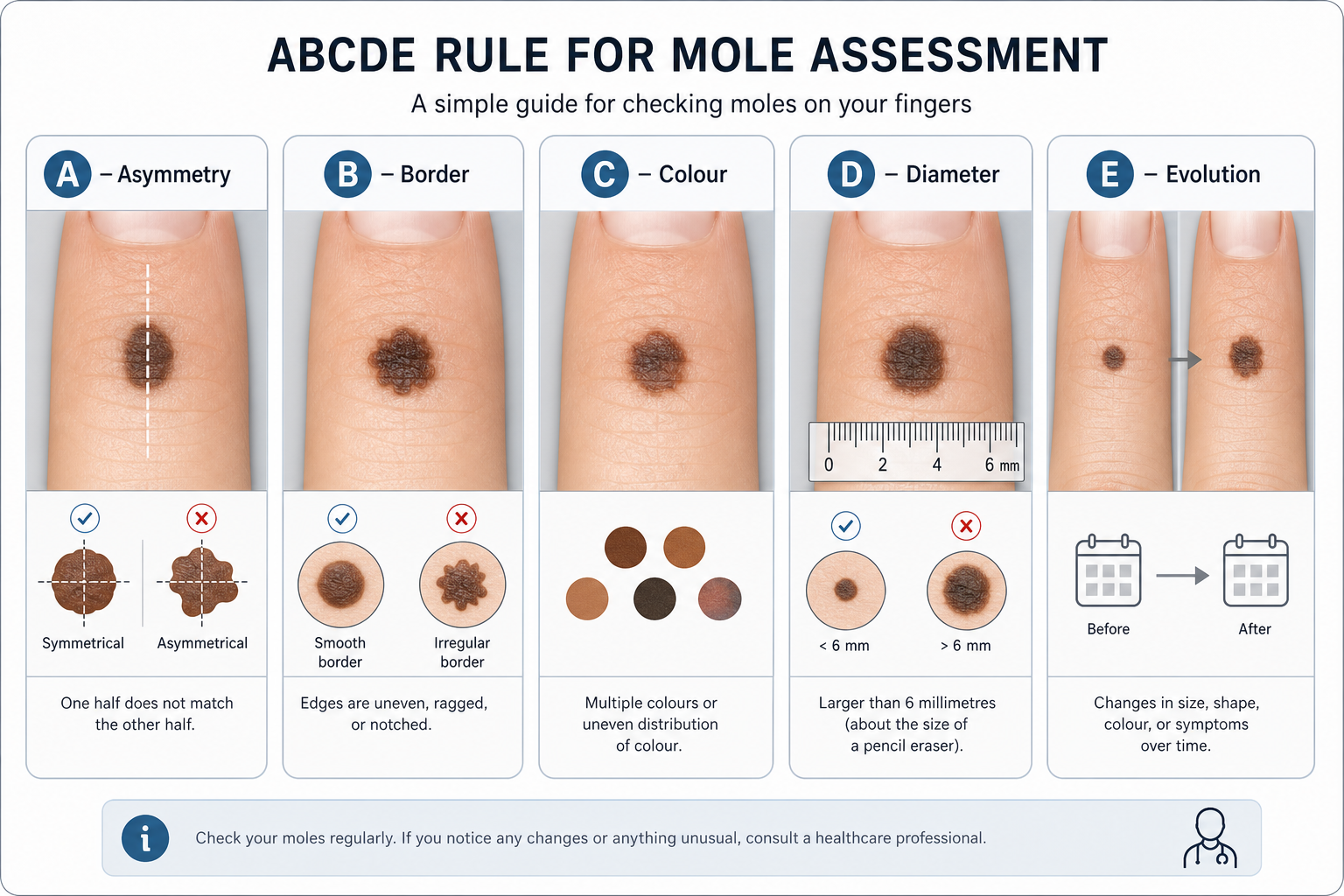
The ABCDE rule is the internationally recognized framework used by dermatologists and taught to patients for self-monitoring of moles. Applying it to a mole on the finger is straightforward, though the small surface area and frequent movement of fingers can make self-examination slightly more challenging than on the trunk or back.
| Letter | Criterion | What to Look For on a Finger Mole |
|---|---|---|
| A | Asymmetry | One half of the mole does not match the other when you draw an imaginary line through the centre |
| B | Border | Edges are ragged, notched, blurred, or irregular rather than smooth and well-defined |
| C | Colour | Color variation within a mole — shades of brown, black, red, white, or blue within the same lesion |
| D | Diameter | Larger than approximately 6 mm (roughly the size of a pencil eraser), though melanomas can be smaller |
| E | Evolution | Any change in size, shape, colour, or texture over weeks or months; new symptoms such as itching, bleeding, or crusting |
The “ugly duckling” sign is a complementary concept: a mole that looks noticeably different from all your other moles — regardless of whether it meets every ABCDE criterion — deserves professional attention. On the fingers, where moles are relatively uncommon compared with the trunk, any lesion that appears distinctly out of place warrants a closer look.
Acral Lentiginous Melanoma: The Finger-Specific Cancer Risk
Acral lentiginous melanoma (ALM) is a subtype of melanoma that develops on acral skin — the palms, soles, and fingers. Unlike the more common superficial spreading melanoma, ALM does not require UV exposure to develop, which means it can arise on the palmar side of the finger and other non-sun-exposed surfaces. This is a clinically important distinction: people who assume that skin cancer only develops on sun-exposed skin may overlook a lesion on the inner surface of a finger or fingertip.
ALM accounts for a small proportion of all melanoma cases overall, but it represents a disproportionately high share of melanoma diagnoses in people with darker skin tones — including those of African, Asian, and Hispanic heritage. Because ALM is less commonly associated with fair skin and sun damage, it is sometimes diagnosed at a later stage, which affects prognosis. Early recognition and prompt biopsy are therefore particularly important. ALM on the finger typically appears as an irregularly shaped, flat, dark brown or black patch that may slowly enlarge over months to years. It can be mistaken for a bruise, a fungal infection, or a benign pigmented lesion.
Subungual Melanoma: When the Concern Is Under or Near the Nail
Subungual melanoma is a form of acral lentiginous melanoma that arises in the nail matrix — the tissue beneath the base of the nail — and is distinct from a mole on the surface of the finger skin. It most commonly affects the thumb or big toe but can occur on any digit. The earliest visible sign is often longitudinal melanonychia: a dark brown or black streak running lengthwise along the nail plate from the base to the tip. This streak originates in the nail matrix and represents pigment produced by abnormal melanocytes.
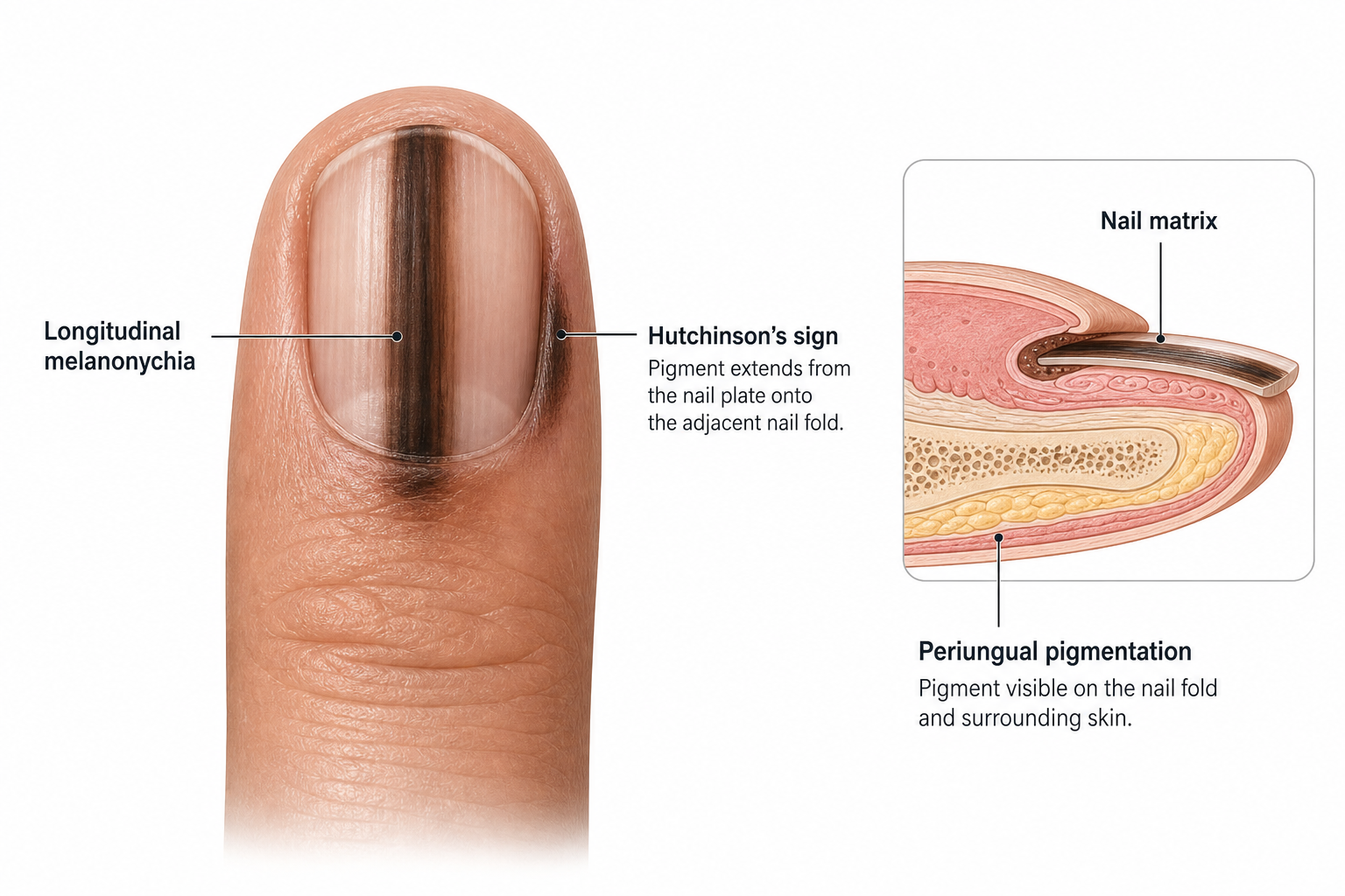
Not every dark nail streak is subungual melanoma — longitudinal melanonychia can also result from nail trauma, certain medications, systemic conditions, or benign melanocyte activation, particularly in people with darker skin tones where it is relatively common. However, features that raise concern include a streak that is wider than 3 mm, has irregular or blurred edges, is darkening or widening over time, or is accompanied by periungual pigmentation — a brownish discolouration spreading from the nail onto the surrounding skin fold. This last sign is known as Hutchinson’s sign and is considered a significant warning indicator that warrants urgent dermatological review. Any new or changing dark streak under the nail should be assessed by a dermatologist rather than monitored at home.
Palmar vs. Dorsal: Why Location on the Finger Matters
The dorsal surface of the fingers — the back of the hand side — receives meaningful UV exposure during everyday activities such as driving, gardening, and outdoor work. Cumulative sun damage on this surface increases the risk of common UV-related skin cancers including basal cell carcinoma and squamous cell carcinoma, as well as the more common UV-associated melanoma subtypes. Applying broad-spectrum sun protection to the backs of the hands is a practical and often overlooked preventive measure.
The palmar side of the finger, by contrast, receives little to no UV exposure. A pigmented lesion on the palmar surface is therefore less likely to be UV-related and more likely to fall into the acral lentiginous melanoma category if it is malignant. This distinction matters because it challenges the assumption that skin cancer is primarily a sun-exposure problem. A mole or dark patch on the inner surface of a finger deserves the same careful assessment as one on the sun-exposed dorsal surface — arguably more so, given that ALM is often caught later.
Risk Factors for a Concerning Mole on the Finger
Several factors may increase the likelihood that a mole on the finger warrants closer attention or more frequent monitoring:
- A new mole after age 30: Most benign nevi develop before the age of 30. A new mole in adulthood — particularly after age 40 or 50 — is more likely to require investigation than one that has been present since childhood.
- Personal or family history of melanoma: A first-degree relative with melanoma, or a personal history of any melanoma, significantly increases lifetime risk.
- Multiple dysplastic nevi: Having ten or more atypical moles is associated with a substantially elevated melanoma risk. People with dysplastic nevus syndrome (also called FAMMM syndrome when familial) benefit from structured surveillance.
- Darker skin tone: While overall melanoma risk is lower in people with darker skin, the proportion of cases that are acral lentiginous melanoma or subungual melanoma is higher, and these are often diagnosed at a more advanced stage.
- Nail trauma or chronic irritation: Repeated trauma to the nail or fingertip has been proposed as a contributing factor in some cases of subungual melanoma, though the evidence is not conclusive.
- Immunosuppression: People who are immunosuppressed — for example, following organ transplantation or due to certain medical conditions — have a higher risk of skin cancers including squamous cell carcinoma on the hands and fingers.
- Cumulative UV exposure on the dorsal hand: Outdoor workers, people who drive frequently without gloves, and those who have had significant sun exposure over a lifetime face higher UV-related risk on the back of the hand.
Warning Signs That Need Prompt Attention
The following changes in a mole on the finger should prompt a dermatologist appointment without delay rather than a period of watchful waiting at home:
- The mole has changed in size, shape, or colour over a period of weeks to months.
- The border has become irregular, notched, or blurred where it was previously smooth.
- There is color variation within a mole — multiple shades of brown, black, red, or white within the same lesion.
- The mole is bleeding, oozing, or crusting without an obvious injury to explain it.
- There is persistent itching, tenderness, or a burning sensation in or around the mole.
- A dark streak has appeared under the nail, particularly if it is widening, darkening, or accompanied by periungual pigmentation.
- The mole looks noticeably different from all your other moles — the “ugly duckling” sign.
- A new pigmented lesion has appeared on the palmar side of the finger or fingertip in an adult over 30.
How Finger Moles Are Diagnosed
A dermatologist will typically begin with a visual examination of the mole in the context of your full skin history, including any family history of melanoma, your skin type, and how long the lesion has been present. Dermoscopy — the use of a handheld dermatoscope that illuminates and magnifies the skin surface — allows the clinician to examine the internal structure of a mole in detail that is not visible to the naked eye. Dermoscopy significantly improves the accuracy of distinguishing benign from potentially malignant lesions and is standard practice in specialist mole assessment.
If the mole has features that cannot be confidently classified as benign by dermoscopy, a skin biopsy will be recommended. A biopsy involves removing all or part of the lesion under local anaesthetic and sending it to a pathology laboratory for microscopic analysis. This is the only definitive way to confirm or exclude melanoma or other skin cancers. For a suspected subungual melanoma, the biopsy involves sampling the nail matrix, which requires a specialist procedure. Mole mapping — systematic full-body photography combined with dermoscopic imaging — may be recommended for people with multiple moles or a high-risk profile, allowing changes to be tracked accurately over time.
Mole Removal Options for Finger Moles
If a biopsy confirms that a mole is benign but the patient wishes to have it removed — for example, because it is repeatedly irritated by rings, tools, or daily hand use — several options may be discussed with a dermatologist or plastic surgeon.
- Surgical excision: The mole is cut out with a scalpel under local anaesthetic and the wound is closed with sutures. Surgical excision is the standard approach for any mole with atypical features, as it provides a complete tissue sample for pathological analysis. On the finger, healing may take longer than on less mobile areas of the body, and the scar may be more noticeable due to the tension placed on the skin during hand movement.
- Shave removal: Suitable only for raised, clearly benign moles, shave removal uses a blade to remove the mole flush with the skin surface without deep excision. It is not appropriate for moles with any atypical features, as it does not provide a full-thickness sample.
- Laser removal: Laser treatment can be used for certain flat, pigmented, benign lesions, but it is generally not recommended for moles that have not been histologically confirmed as benign, as it destroys tissue rather than preserving it for analysis.
Post-removal care on the finger requires particular attention. Fingers are constantly in motion, exposed to water, and at risk of contact with surfaces that could introduce infection. Keeping the wound covered, dry, and protected — and avoiding activities that stretch or stress the healing skin — is important for minimising scarring and reducing infection risk. Your treating clinician will provide specific aftercare instructions based on the removal method used.
At-Home Self-Monitoring: A Practical Approach
Monitoring a mole on the finger at home is straightforward with a consistent approach. The goal is not to diagnose a mole yourself but to detect changes early enough to seek professional assessment promptly.
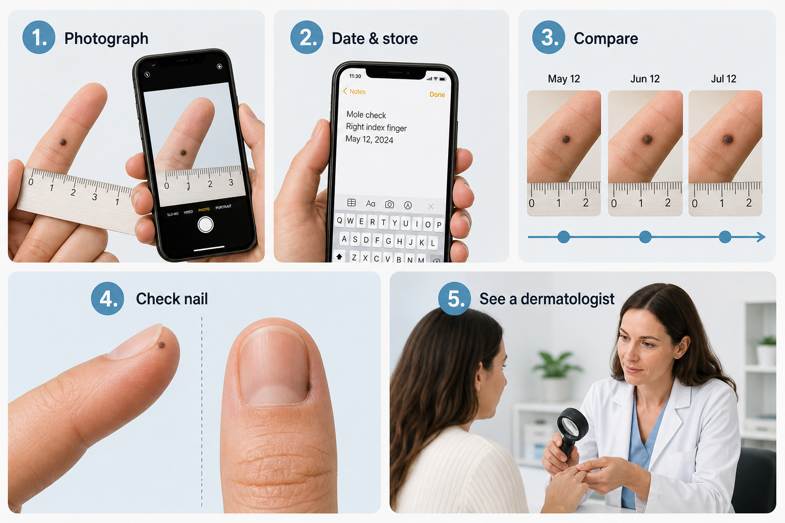
- Photograph the mole in good natural light using a smartphone camera. Take one image of the full finger for context and one close-up of the mole itself. Include a ruler or coin in the frame to provide a size reference.
- Note the date and store the images in a dedicated folder so you can compare them accurately over time.
- Repeat the photograph every 4–8 weeks if you are monitoring a mole that has been assessed as benign but that you want to track. Compare each new image with the previous one rather than relying on memory.
- Check the periungual area and nail surface at the same time, looking for any new dark streaks or changes in existing ones.
- Note any symptoms — itching, tenderness, bleeding, or crusting — even if the mole looks unchanged visually.
- Seek a dermatologist appointment if you notice any change between photographs, any new symptom, or any lesion that simply concerns you. Self-monitoring is a supplement to professional assessment, not a replacement for it.
Fingers are uniquely prone to friction from tools, sports equipment, and jewelry, and to chemical exposure from cleaning products and solvents. Irritation from these sources can cause a benign mole to become temporarily inflamed, itchy, or slightly raised. If a mole on the finger appears irritated after a specific activity or chemical exposure, it is worth noting the context — but persistent symptoms that do not resolve within a few days still warrant professional review.
Frequently Asked Questions
Is a mole on the finger dangerous?
Most moles on the finger are benign and pose no health risk. However, the fingers and nail area are sites where acral lentiginous melanoma and subungual melanoma can develop, so any mole that is new, changing, or has irregular features should be assessed by a dermatologist rather than assumed to be harmless.
Can I remove a mole on my finger at home?
No. At-home mole removal is not safe and is strongly discouraged by dermatologists. Removing a mole without professional assessment means that any potentially malignant tissue is destroyed without being examined, delaying diagnosis. Home removal methods also carry risks of infection, scarring, and incomplete removal.
What does a dark streak under my fingernail mean?
A dark streak running lengthwise under the nail is called longitudinal melanonychia. It has several possible causes, including benign melanocyte activation (common in darker skin tones), nail trauma, certain medications, and — less commonly — subungual melanoma. A new, widening, or irregular streak, or one accompanied by pigment spreading onto the surrounding skin (Hutchinson’s sign), should be assessed by a dermatologist promptly.
Do finger moles need to be removed?
A benign mole on the finger does not need to be removed unless it is causing persistent irritation, discomfort, or cosmetic concern. If a mole has atypical features or a biopsy suggests dysplasia or malignancy, removal is recommended. The decision should be made in consultation with a dermatologist based on the individual mole’s characteristics.
Are people with darker skin tones at risk from finger moles?
People with darker skin tones have a lower overall risk of UV-related melanoma but a higher proportion of acral lentiginous melanoma and subungual melanoma relative to other melanoma subtypes. These cancers are not related to sun exposure and can develop on the palmar side of the finger and under the nail. Awareness of these specific risks is important across all skin tones.
How often should I have a mole on my finger checked?
If a dermatologist has assessed a finger mole and confirmed it is benign, they will advise on an appropriate monitoring interval — typically annually for people with a higher-risk profile, or less frequently for those with a single stable mole and no risk factors. Any change between scheduled appointments should prompt an earlier review rather than waiting for the next routine check.
Can a mole on the finger be a sign of something other than skin cancer?
Yes. Most finger moles are entirely benign nevi with no systemic significance. Occasionally, pigmented lesions on the fingers can be associated with certain systemic conditions, but this is uncommon. If you have concerns about a lesion that does not look like a typical mole, a dermatologist can assess it and arrange any further investigations if needed.
When to Act on a Mole on Your Finger
The practical takeaway is straightforward: most moles on the finger are benign and will remain so throughout a person’s lifetime. Regular self-monitoring using the ABCDE rule and the “ugly duckling” sign, combined with periodic professional skin examinations, is the most effective strategy for catching any change early. The threshold for seeking a dermatologist’s opinion should be low — a consultation is a brief, non-invasive appointment, and dermoscopy can usually provide reassurance or identify a concern quickly. If a skin biopsy is needed, it is a minor procedure performed under local anaesthetic with a short recovery period.
People with a personal or family history of melanoma, multiple dysplastic nevi, or a high-risk occupational or recreational sun exposure history on the dorsal hand should discuss a structured mole mapping programme with a dermatologist. For everyone else, the key message is not to ignore a mole on the finger that is new, changing, or simply looks different — early assessment is always preferable to delayed diagnosis, and in the case of melanoma, timing genuinely affects outcomes.