Laser for Dark Spots: Which Pigment-Targeting Option Is Right for You?
Dark spots — whether caused by sun exposure, hormonal changes, acne, or ageing — are among the most common reasons people seek professional skin treatment. Laser therapy has become one of the most clinically established approaches for reducing hyperpigmentation, but the term “laser for dark spots” covers a wide range of technologies, each with different mechanisms, suitable conditions, and safety profiles. Understanding the differences before booking a consultation can help you ask better questions and set realistic expectations.
Key Takeaways
- No single laser treats every type of dark spot equally well — the right choice depends on the pigmentation condition, its depth in the skin, and your Fitzpatrick skin type.
- Picosecond and Q-switched lasers are among the most widely used for discrete pigmented lesions, while fractional non-ablative devices are often preferred for diffuse or mixed pigmentation.
- Melasma requires a particularly cautious approach; aggressive laser settings can worsen pigmentation rather than improve it.
- Skin of color carries a higher risk of paradoxical post-inflammatory hyperpigmentation if the wrong device or settings are used — specialist assessment is essential.
- Laser treatment addresses existing pigment but does not prevent new spots from forming; broad-spectrum photoprotection and maintenance sessions are central to long-term results.
How Lasers Target Pigment in the Skin
The scientific principle underlying laser pigment treatment is selective photothermolysis — the delivery of a specific wavelength of light that is preferentially absorbed by a target molecule, called a chromophore, while leaving surrounding tissue largely unaffected. In pigmentation treatment, the primary chromophore is melanin, the pigment produced by melanocyte cells. When laser energy is absorbed by melanin-rich cells, it generates heat that disrupts or fragments the pigment granules. The body’s lymphatic system then gradually clears the fragmented melanin, causing the spot to fade over days to weeks.
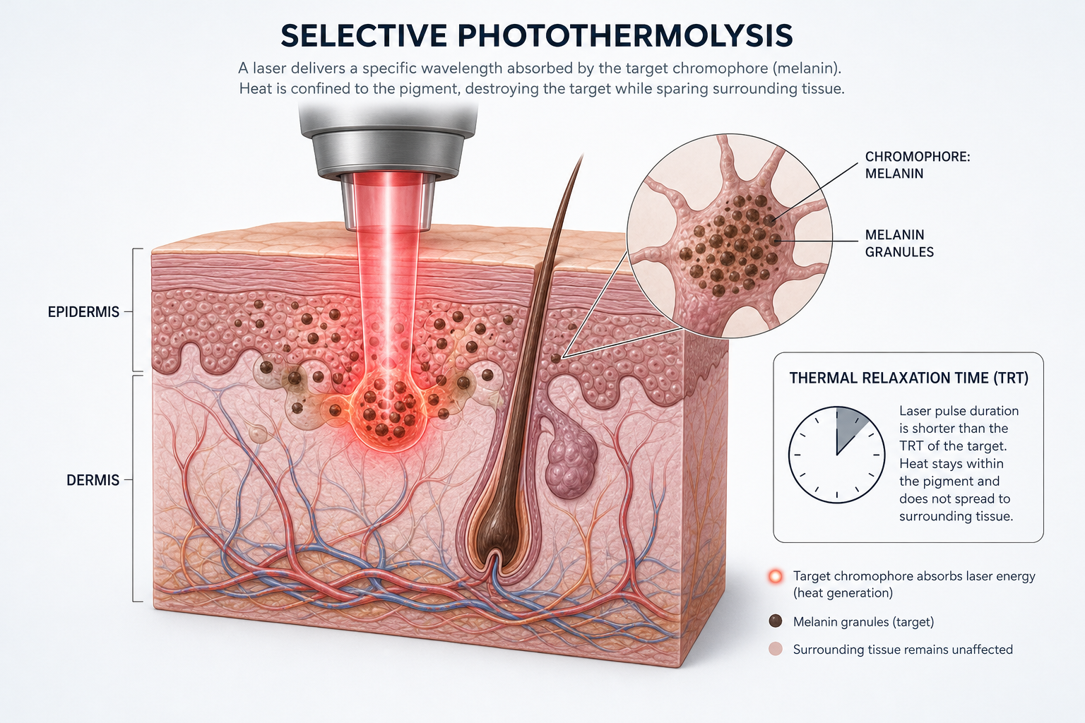
A critical concept in chromophore targeting is thermal relaxation time — the time it takes for a heated target to dissipate half of its absorbed heat into surrounding tissue. To destroy the target without damaging adjacent structures, the laser pulse must be shorter than the thermal relaxation time of the melanin granule. This is why pulse duration matters: very short pulses, such as those produced by picosecond lasers, can shatter pigment mechanically through a photoacoustic effect rather than purely through heat, which may reduce collateral thermal injury.
Equally important is pigment depth. Melanin can sit in the outer skin layer (epidermis), deeper in the dermis, or in both layers simultaneously. Shorter wavelengths, such as 532 nm, are absorbed more superficially and are effective for epidermal pigment. Longer wavelengths, such as 1064 nm, penetrate deeper and can reach dermal melanin. Choosing a laser without considering whether the pigment is epidermal or dermal is one of the most common reasons for suboptimal results.
Wavelength and Pigment Depth: A Practical Overview
Different laser wavelengths interact with melanin at different skin depths, which directly influences which conditions each device can address and which skin tones it can be used on safely.
| Wavelength | Laser / Device | Primary Pigment Depth | Common Uses | Skin Tone Suitability |
|---|---|---|---|---|
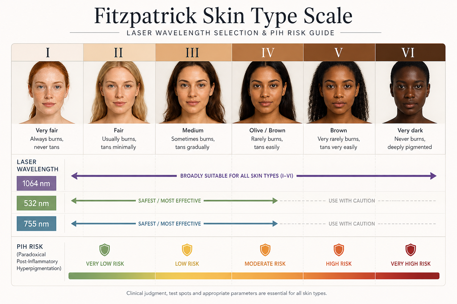
| 532 nm | Q-switched Nd:YAG (KTP), picosecond 532 | Epidermal | Solar lentigines, freckles, superficial sun spots | Fitzpatrick I–III; caution in darker types |
| 694 nm | Ruby laser | Epidermal to superficial dermal | Lentigines, freckles, some tattoo pigment | Fitzpatrick I–III |
| 755 nm | Alexandrite laser, picosecond 755 (PicoSure) | Epidermal to mid-dermal | Sun spots, age spots, some melasma | Fitzpatrick I–IV with care |
| 1064 nm | Q-switched Nd:YAG, picosecond 1064 (PicoWay) | Dermal and epidermal | Dermal melasma, Hori’s nevus, nevus of Ota, PIH in darker skin | Fitzpatrick I–VI (most versatile) |
| 1550 nm | Fraxel Restore (erbium fibre) | Mid to deep dermis (fractional) | Diffuse photoaging, acne scars with PIH, uneven skin tone | Fitzpatrick I–IV; careful patient selection for V–VI |
| 1927 nm | Fraxel DUAL, MOXI, thulium laser | Superficial dermis (fractional) | Epidermal pigmentation, diffuse sun damage, early photoaging | Fitzpatrick I–IV; emerging evidence for V–VI at low settings |
This wavelength-to-depth relationship explains why a device that works well for fair-skinned patients with solar lentigines may be entirely inappropriate for a patient with deeper skin tone and dermal melasma. A pigment-safe wavelength for one condition is not automatically safe or effective for another.
Main Laser Types Used for Dark Spots
Q-Switched Lasers
Q-switched lasers deliver very short, high-energy pulses — typically in the nanosecond range — that generate rapid thermal expansion within melanin granules, fragmenting them. The Q-switched Nd:YAG laser, available at both 1064 nm and 532 nm, has a long clinical track record for treating solar lentigines, age spots, and certain dermal pigmentation conditions. At 1064 nm with low-fluence settings, Q-switched Nd:YAG is one of the few laser options with reasonable evidence for use in darker Fitzpatrick skin types, including for conditions such as Hori’s nevus and nevus of Ota. Multiple sessions are typically required, and results depend heavily on the specific condition and skin type.
Picosecond Lasers
Picosecond lasers, such as PicoSure (755 nm) and PicoWay (532 nm and 1064 nm), deliver pulses in the trillionths-of-a-second range — significantly shorter than Q-switched nanosecond devices. This ultrashort picosecond pulse duration produces a stronger photoacoustic (pressure-wave) effect, which can shatter melanin granules into smaller particles that are more readily cleared by the body. Clinical studies suggest picosecond lasers may achieve comparable or improved pigment clearance with fewer sessions and potentially less thermal injury compared with nanosecond Q-switched devices, though evidence continues to develop and results vary by condition. Picosecond platforms are increasingly used for solar lentigines, post-inflammatory hyperpigmentation, and selected cases of melasma.
Fractional Non-Ablative Lasers
Fractional lasers, including Fraxel (1550 nm and 1927 nm), MOXI, and Clear + Brilliant, treat thousands of microscopic columns of skin while leaving surrounding tissue intact. This fractional non-ablative approach stimulates collagen production and promotes epidermal turnover, which can gradually improve diffuse pigmentation, uneven skin tone, and photoaging. Because only a fraction of the skin surface is treated at each session, recovery is generally faster than with fully ablative resurfacing. These devices are particularly useful when pigmentation is widespread rather than confined to discrete spots, and when skin rejuvenation is also a goal. The 1927 nm thulium wavelength targets the superficial dermis and has shown promise for epidermal pigmentation, including in some studies involving patients with darker skin tones at appropriately conservative settings.
IPL (Intense Pulsed Light)
Intense Pulsed Light is not technically a laser — it emits a broad spectrum of light wavelengths filtered to target specific chromophores. IPL can be effective for diffuse sun damage, solar lentigines, and redness in lighter skin types, and is often used as part of a photorejuvenation programme. However, because IPL delivers a range of wavelengths rather than a single precise wavelength, it offers less selective chromophore targeting than a true laser. IPL is generally not recommended for darker skin tones or for conditions such as melasma, where non-specific heating can trigger rebound pigmentation.
Ablative Fractional Lasers
Fractional CO2 and erbium:YAG lasers remove microscopic columns of skin tissue entirely, stimulating significant collagen remodelling and skin resurfacing. These devices can address pigmentation alongside textural concerns such as acne scars and deep wrinkles, but they carry greater downtime — typically one to two weeks of visible recovery — and a higher risk of post-inflammatory hyperpigmentation in patients with darker skin tones. Ablative fractional resurfacing is generally reserved for patients with Fitzpatrick types I–III and is not a first-line choice for isolated pigmentation concerns.
Matching the Laser to the Pigmentation Condition
Solar Lentigines and Age Spots
Solar lentigines — flat, well-defined brown spots caused by cumulative UV exposure — are among the most straightforward pigmentation conditions to treat with laser. They contain excess melanin concentrated in the epidermis, making them responsive to Q-switched and picosecond devices at 532 nm or 755 nm. Clearance is often achievable in one to three sessions for discrete lesions, though new spots can develop with continued sun exposure. Consistent broad-spectrum photoprotection after treatment is essential to prevent recurrence.
Melasma
Melasma is a hormonally influenced, often chronic pigmentation condition that can involve both epidermal and dermal melanin — and sometimes both simultaneously. It is notoriously prone to rebound pigmentation, meaning that aggressive laser treatment can trigger an inflammatory response that stimulates melanocytes to produce more pigment, worsening the condition. For this reason, melasma requires a fundamentally different approach than discrete sun spots. Low-fluence Q-switched Nd:YAG protocols, sometimes called “laser toning,” have been used for melasma in Asian skin types, though evidence on long-term efficacy and recurrence rates remains mixed. Picosecond lasers are increasingly studied for melasma, with some trials showing meaningful improvement, particularly for epidermal-predominant cases. Any laser treatment for melasma should be combined with strict photoprotection, and often with topical agents such as tranexamic acid, hydroquinone, or azelaic acid, as part of a structured combination laser protocol. Patients should be counselled that melasma is a chronic condition and that recurrence is common, particularly with sun exposure or hormonal changes.
Post-Inflammatory Hyperpigmentation (PIH)
Post-inflammatory hyperpigmentation develops after skin injury or inflammation — commonly following acne, eczema, or a previous cosmetic procedure. PIH is predominantly epidermal in lighter skin types but can extend into the dermis in patients with melanin-rich skin. Laser treatment for PIH requires careful patient selection: the same inflammatory stimulus that caused the original PIH can be triggered by laser energy itself, leading to paradoxical post-inflammatory hyperpigmentation — a worsening of the very condition being treated. Conservative settings, appropriate wavelength selection, and a thorough pre-treatment assessment are essential. Fractional non-ablative devices at low energy levels and long-wavelength Q-switched or picosecond lasers are generally preferred. Topical pre-treatment with agents such as hydroquinone or kojic acid for several weeks before laser can help reduce the risk of PIH in susceptible patients.
Freckles
Freckles (ephelides) are superficial, epidermal pigmentation spots that respond well to laser treatment, particularly with Q-switched or picosecond devices at 532 nm or 755 nm. Because freckles are genetically determined and influenced by UV exposure, they tend to return with sun exposure even after successful treatment. Patients should understand that laser can significantly reduce freckle visibility but is unlikely to prevent new ones from appearing without diligent photoprotection.
Acne-Related Dark Marks
Dark marks left by acne are a form of post-inflammatory hyperpigmentation and are treated using the same principles. Fractional non-ablative lasers are often useful here because they can address both the pigmentation and any associated textural scarring simultaneously. The 1927 nm thulium wavelength and picosecond devices with fractional handpieces have shown utility in this context. As with other PIH, the risk of paradoxical darkening must be managed through appropriate device selection and settings.
Laser Safety for Skin of Color
Patients with darker Fitzpatrick skin types (IV through VI) face a meaningfully higher risk of adverse effects from laser pigmentation treatment, including paradoxical post-inflammatory hyperpigmentation, hypopigmentation (permanent lightening of treated skin), and scarring. These risks arise because the epidermis in darker skin contains more melanin, which competes with the target pigment for laser energy absorption. When epidermal melanin absorbs excessive energy, it can be damaged, triggering an inflammatory cascade that stimulates further pigment production — the opposite of the intended outcome.
For patients with skin of color, the safest laser options are generally those using longer wavelengths with lower fluence settings. The 1064 nm Nd:YAG — both Q-switched and picosecond — is the most widely validated wavelength for darker skin tones because its longer wavelength is less aggressively absorbed by epidermal melanin, allowing it to reach deeper targets with reduced surface risk. Emerging research on 785 nm picosecond devices and 1927 nm fractional thulium lasers at conservative settings also shows promise for darker phototypes, though evidence is still developing. A test spot on a small, inconspicuous area before full treatment is a standard precaution in this patient group. Choosing a practitioner with documented experience treating skin of color is as important as choosing the right device.
What Happens During a Laser Pigmentation Treatment
A typical laser session for dark spots begins with a thorough skin assessment, including a review of the pigmentation type, distribution, and likely depth. Photographs are usually taken for comparison. The skin is cleansed and, depending on the device and patient sensitivity, a topical anaesthetic cream may be applied and left for 20 to 45 minutes before treatment begins.
During the session, the practitioner passes the laser handpiece over the treatment area, delivering pulses of light to the pigmented zones. Patients commonly describe the sensation as a snapping or stinging feeling, similar to a rubber band flicking against the skin. The duration of a session varies from a few minutes for a small area to 30 minutes or more for a full face or larger body area.
Immediately after treatment, the skin may appear red and slightly swollen. Treated spots often darken initially — a normal response as the fragmented melanin rises toward the skin surface before being shed. This temporary darkening, sometimes called the “purge” effect, can alarm patients who are not forewarned. Over the following one to three weeks, the darkened areas gradually flake away or fade, revealing lighter skin beneath. The timeline varies by device, energy level, and individual healing response.
Recovery, Aftercare, and Maintenance
Recovery after non-ablative laser sessions is generally mild: redness and mild swelling typically resolve within 24 to 72 hours. Fractional non-ablative treatments may cause a sandpaper-like texture and bronzed appearance for three to seven days as the treated micro-columns shed. Ablative fractional treatments involve more significant downtime, with redness, crusting, and sensitivity lasting one to two weeks.
Aftercare consistently recommended across clinical settings includes:
- Applying a gentle, fragrance-free moisturiser to support the skin barrier during healing.
- Using a broad-spectrum photoprotection product of SPF 30 or higher every morning, reapplied throughout the day — this is non-negotiable after any pigmentation laser treatment.
- Avoiding direct sun exposure, particularly during peak UV hours, for at least four to six weeks post-treatment.
- Avoiding active skincare ingredients such as retinoids, exfoliating acids, and vitamin C serums until the skin has fully healed, typically one to two weeks.
- Not picking or rubbing treated areas, as this can cause PIH or scarring.
Long-term maintenance is an important and often underemphasised aspect of laser pigmentation treatment. UV exposure is the primary driver of new melanin production, meaning that without consistent sun protection, new dark spots will form regardless of how successful the initial treatment was. Many patients benefit from periodic maintenance sessions — typically once or twice a year — combined with a daily topical regimen that may include ingredients such as tranexamic acid, niacinamide, or retinoids to slow pigment recurrence.
How Many Sessions Are Typically Needed?
The number of sessions required depends on the pigmentation condition, its severity, the device used, and individual response. As a general guide:
- Discrete solar lentigines and age spots: Often 1–3 sessions with Q-switched or picosecond lasers.
- Diffuse sun damage and photoaging: Typically 3–5 sessions with fractional non-ablative devices, spaced four to six weeks apart.
- Melasma: Variable; may require 4–8 or more sessions with conservative settings, and ongoing maintenance. Results are less predictable than for discrete lesions.
- Post-inflammatory hyperpigmentation: Typically 3–6 sessions, depending on depth and skin tone.
Sessions are usually spaced four to eight weeks apart to allow the skin to heal and the body to clear fragmented pigment before the next treatment. Rushing the interval between sessions increases the risk of cumulative inflammation and adverse effects.
Realistic Costs to Consider
Laser pigmentation treatment is not covered by health insurance in most countries, as it is considered a cosmetic procedure. Costs vary considerably by device, clinic location, practitioner experience, and the size of the treatment area. As a general indication, individual sessions for discrete spot treatment may range from approximately £100 to £400 per session, while full-face fractional treatments can range from £300 to £800 or more per session. For conditions such as melasma that require multiple sessions and ongoing maintenance, the total investment over time can be substantial. Requesting a full treatment plan with estimated total cost — not just a per-session price — during the consultation gives a more accurate picture of the financial commitment involved.
Questions to Ask Before Booking a Laser Treatment
A well-informed consultation is the most important step before any laser pigmentation treatment. Consider asking your practitioner:
- What type of pigmentation do I have, and is it epidermal, dermal, or mixed?
- Which laser or device do you recommend for my specific condition and skin tone, and why?
- What is the risk of paradoxical post-inflammatory hyperpigmentation for someone with my Fitzpatrick skin type?
- How many sessions are realistically needed, and what is the expected interval between them?
- What does the full treatment plan look like, including aftercare and maintenance?
- What are the possible side effects, and how would they be managed if they occurred?
- Will you perform a test spot before treating the full area?
- What topical treatments should I use before and after laser to optimise results and reduce risk?
Frequently Asked Questions
Can laser permanently remove dark spots?
Laser treatment can significantly reduce or eliminate existing dark spots, but it does not prevent new ones from forming. Solar lentigines treated with laser may not return if sun exposure is consistently avoided, but UV exposure, hormonal changes, and skin inflammation can all trigger new pigment production. Long-term results depend heavily on sun protection habits and, for conditions like melasma, ongoing management.
Is laser safe for dark skin tones?
Laser treatment can be performed safely on darker skin tones, but it requires careful device selection, appropriate wavelengths, and conservative energy settings. The 1064 nm Nd:YAG laser — in both Q-switched and picosecond formats — has the strongest evidence base for use in Fitzpatrick types IV through VI. Choosing a practitioner with specific experience in treating skin of color is essential to minimise the risk of paradoxical hyperpigmentation or hypopigmentation.
Why do dark spots look darker immediately after laser treatment?
Temporary darkening of treated spots after laser is a normal and expected response. The laser fragments melanin granules, which then migrate toward the skin surface before being shed through natural skin turnover. This process typically takes one to three weeks. Patients who are not warned about this effect sometimes mistake it for worsening and discontinue treatment prematurely.
What is the difference between laser and IPL for dark spots?
A laser emits a single, precise wavelength of light, allowing highly selective chromophore targeting. IPL emits a broad spectrum of wavelengths filtered to a range, which makes it less precise but useful for treating multiple concerns simultaneously, such as pigmentation and redness. For discrete, well-defined dark spots, a targeted laser generally offers more controlled treatment. IPL is better suited to diffuse sun damage in lighter skin types and is not recommended for melasma or darker skin tones.
Can I combine laser with topical treatments?
Yes, and combination approaches are often recommended. Topical agents such as hydroquinone, tranexamic acid, kojic acid, azelaic acid, and retinoids can be used before laser to reduce melanocyte activity and lower the risk of PIH, and after laser to maintain results and slow pigment recurrence. Your practitioner should advise on which topicals are appropriate for your skin type and condition, and when to pause them around treatment sessions.
How long after laser treatment can I go in the sun?
Sun exposure should be minimised for at least four to six weeks after laser pigmentation treatment, and broad-spectrum SPF 30 or higher should be applied daily from the day after treatment onwards. UV exposure during the healing phase is one of the most common causes of post-treatment PIH and can significantly undermine results.
Choosing the Right Laser Approach for Your Dark Spots
Laser treatment for dark spots is not a one-size-fits-all solution. The most appropriate device depends on the specific pigmentation condition, whether the melanin is epidermal or dermal, your Fitzpatrick skin type, and your tolerance for downtime and treatment frequency. Discrete solar lentigines in lighter skin tones are among the most straightforward conditions to treat, often responding well to Q-switched or picosecond lasers in a small number of sessions. Melasma and post-inflammatory hyperpigmentation in darker skin tones are considerably more complex, requiring conservative protocols, combination topical strategies, and realistic expectations about recurrence.
The most important step is a thorough consultation with a practitioner who can accurately diagnose your pigmentation type, assess your skin tone, and recommend a treatment plan tailored to your individual circumstances — rather than applying a generic protocol. If you are considering laser for dark spots, prioritise practitioners with demonstrable experience in pigmentation treatment and, if relevant, in treating skin of color. A well-planned approach that combines the right laser technology with appropriate aftercare and sun protection gives the best chance of meaningful, lasting improvement.