Scabby Mole Causes: What You Need to Know
A mole that develops a scab, crust, or rough surface can be unsettling to notice. While many cases have straightforward explanations such as minor trauma or irritation, a scabby mole should never be dismissed without consideration, because changes to a mole’s surface can occasionally indicate something that warrants professional assessment. This article explains the most common causes of a scabby mole, the warning signs that require prompt medical attention, and what you can expect if you decide to have a mole evaluated or removed.
Key Takeaways
- A mole that repeatedly scabs, bleeds without obvious cause, or fails to heal should be assessed by a qualified clinician promptly.
- Many scabby moles result from benign causes such as friction, accidental scratching, or minor skin conditions, but this cannot be assumed without examination.
- The ABCDE criteria — Asymmetry, Border, Colour, Diameter, Evolution — are a useful self-monitoring framework, but they do not replace professional dermoscopic evaluation.
- Dermatoscopy and, where indicated, biopsy are the standard clinical tools used to assess a changing mole accurately.
- Early assessment of any suspicious mole change is always preferable to a wait-and-see approach.
What Does a Scabby Mole Actually Mean?
A scab forms when the skin’s surface is disrupted and the body’s natural clotting and repair process creates a protective crust over the wound. When this happens on or around a mole, it means the skin overlying or adjacent to the mole has been broken in some way. The key clinical question is whether that disruption has an obvious, innocent explanation — such as catching the mole on clothing — or whether the mole itself is changing in a way that has caused the surface to break down without any external trigger.
Moles, known medically as melanocytic naevi, are clusters of pigment-producing cells called melanocytes. Most moles are entirely benign and remain stable throughout a person’s life. However, some moles can undergo changes over time, and surface changes such as crusting, ulceration, or bleeding are among the features that clinicians take seriously during skin assessments. The presence of a scab alone does not confirm a diagnosis in either direction, which is precisely why professional evaluation matters.
Common Benign Causes of a Scabby Mole
The majority of moles that develop a scab or crust do so for straightforward, non-threatening reasons. Understanding these causes can help you assess whether there is an obvious explanation for the change you have noticed.
Friction and Repeated Irritation
Moles located in areas where clothing, jewellery, or skin rubs repeatedly are particularly prone to surface disruption. Common friction sites include the bra line, collar area, waistband, inner thighs, and underarms. Repeated rubbing gradually wears down the surface of the mole, causing minor abrasions that then scab over as they heal. If the friction continues, the cycle of irritation and scabbing may repeat. This is one of the most frequent benign explanations for a scabby mole, and it is also one of the clearest reasons why moles in high-friction areas are often considered for removal.
Accidental Scratching or Trauma
Moles can be caught accidentally during shaving, scratched during sleep, or knocked during everyday activities. Because moles are slightly raised in many cases, they are more exposed to incidental contact than the surrounding flat skin. A single traumatic event can break the surface and produce a scab that heals within one to two weeks in most cases. If you can clearly recall an incident that caused the injury and the mole heals completely without further change, this is generally reassuring — though any doubt should still prompt a professional check.
Eczema, Psoriasis, and Other Skin Conditions
Inflammatory skin conditions such as eczema or psoriasis can affect the skin overlying a mole just as they affect the surrounding skin. When these conditions cause itching, the mole may be scratched repeatedly, leading to surface breakdown and crusting. Similarly, seborrhoeic dermatitis — a common condition causing flaky, irritated skin — can affect moles, particularly on the scalp or face. In these situations, the scabbing is a consequence of the underlying skin condition rather than a change within the mole itself, though distinguishing between the two still requires clinical assessment.
Seborrhoeic Keratoses Mistaken for Moles
Seborrhoeic keratoses are extremely common benign skin growths that often have a rough, warty, or crusted surface. They are frequently mistaken for moles, particularly as they can be pigmented and may appear similar to a mole at first glance. Seborrhoeic keratoses naturally develop a scabby or flaky texture as part of their normal appearance, and they may also catch on clothing and bleed. A clinician can usually distinguish a seborrhoeic keratosis from a true melanocytic naevus using dermoscopy, which is a non-invasive magnified examination of the skin.
Insect Bites and Minor Infections
An insect bite on or immediately adjacent to a mole can cause localised swelling, itching, and subsequent scabbing that may appear to involve the mole itself. Similarly, a minor bacterial infection of a hair follicle near a mole can produce crusting. These causes are usually accompanied by other signs such as redness, warmth, or a clear history of a bite, and they typically resolve within a short period.
When a Scabby Mole May Indicate Something More Serious
While benign causes account for many cases, there are specific circumstances in which a scabby or crusting mole requires urgent professional evaluation. The concern in these situations is the possibility of melanoma — a form of skin cancer arising from melanocytes — or other skin cancers such as basal cell carcinoma or squamous cell carcinoma, which can also develop within or adjacent to pigmented lesions.
Spontaneous Bleeding or Crusting Without a Clear Cause
A mole that bleeds or develops a crust without any identifiable trauma is a significant warning sign. Healthy, stable moles do not spontaneously bleed. When a mole bleeds or scabs without an obvious external cause, it may indicate that the cells within the mole are proliferating abnormally, disrupting the overlying skin from within. This symptom should prompt a medical appointment without delay rather than a period of observation at home.
A Scab That Does Not Heal
Normal skin wounds, including those on or near a mole caused by friction or trauma, typically heal within one to two weeks. A scab that persists beyond this timeframe, repeatedly reforms after appearing to heal, or is accompanied by an enlarging or changing mole beneath it warrants professional assessment. Non-healing skin lesions are a recognised feature of certain skin cancers, including basal cell carcinoma, which can sometimes appear as a pearly or translucent lesion with a crusted centre.
Changes Alongside the Scabbing
A scabby mole becomes more concerning when the scabbing is accompanied by other changes to the mole itself. These include an increase in size, a change in shape or border regularity, the development of multiple colours within the same mole, or the appearance of new satellite lesions nearby. The ABCDE framework — Asymmetry, Border irregularity, Colour variation, Diameter greater than 6 mm, and Evolution or change over time — is a widely used patient-facing tool for identifying moles that may need professional review. However, it is important to understand that not all melanomas display every ABCDE feature, and some benign moles may display one or two of these characteristics. The framework is a prompt to seek assessment, not a diagnostic tool.
Moles in High-Risk Individuals
Certain individuals carry a higher baseline risk of developing melanoma, and any change in a mole — including scabbing — should be assessed more promptly in these groups. Risk factors for melanoma include a personal or family history of melanoma, a large number of moles (typically more than 50), a history of significant sun exposure or sunbed use, fair skin that burns easily, a history of blistering sunburn particularly in childhood, and a weakened immune system. People in these groups are generally advised to have regular skin surveillance and to seek assessment for any new or changing mole feature without delay.
The ABCDE Criteria: A Practical Self-Monitoring Guide
The ABCDE criteria provide a structured way for patients to monitor their own moles between professional skin checks. Each letter represents a feature that, when present or changing, suggests a mole may benefit from clinical review.
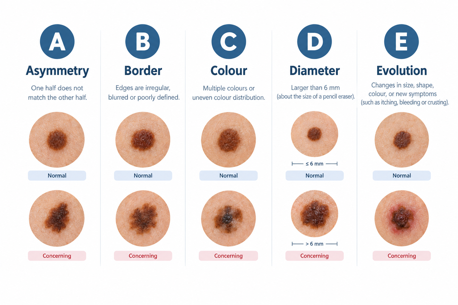
- Asymmetry: One half of the mole does not match the other half in shape.
- Border: The edges of the mole are irregular, ragged, notched, or blurred rather than smooth and well-defined.
- Colour: The mole contains more than one shade of brown, black, red, white, or blue rather than a uniform single colour.
- Diameter: The mole is larger than approximately 6 mm across, roughly the size of a pencil eraser, though melanomas can be smaller.
- Evolution: The mole has changed in size, shape, colour, or surface texture, or has developed new symptoms such as itching, bleeding, or crusting.
A scabby mole falls under the Evolution category of this framework. Even if the other four criteria appear normal, a mole that has developed a new scab or surface change — particularly without a clear cause — satisfies the E criterion and should be assessed by a clinician.
How a Scabby Mole Is Assessed Clinically
When you present to a clinician with a scabby or changing mole, the assessment typically follows a structured process designed to determine whether the mole requires further investigation or treatment.
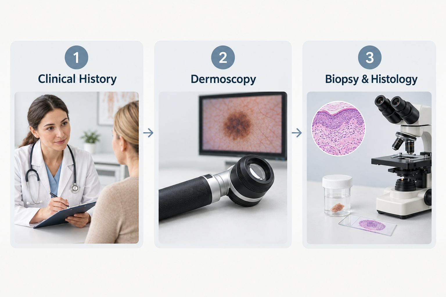
Clinical History
The clinician will ask about how long the scabbing has been present, whether there was any identifiable cause such as trauma or friction, whether the mole has changed in other ways, and about your personal and family history of skin cancer. This history helps to contextualise the physical findings and identify any relevant risk factors.
Dermoscopy
Dermoscopy — also called dermatoscopy — is a non-invasive examination technique using a handheld device that illuminates and magnifies the skin surface. It allows a trained clinician to examine the internal structure of a mole in detail that is not visible to the naked eye. Dermoscopy significantly improves the accuracy of mole assessment compared with naked-eye examination alone and is considered the standard of care for evaluating pigmented skin lesions. It can help distinguish benign moles, seborrhoeic keratoses, and other benign lesions from those that require biopsy.
Biopsy and Histological Examination
When dermoscopy reveals features that cannot be confidently classified as benign, or when a mole has characteristics that raise clinical concern, a biopsy is performed. This involves removing all or part of the mole under local anaesthetic and sending the tissue to a pathology laboratory for microscopic examination. Histological analysis of the tissue sample is the definitive method for diagnosing or excluding melanoma and other skin cancers. The result of a biopsy determines whether further treatment — such as wider surgical excision — is required.
Mole Removal: When and Why It May Be Recommended
Mole removal may be recommended for clinical reasons — when a mole is suspicious or has been confirmed as abnormal — or for practical reasons, such as persistent irritation from friction in a high-movement area. In some cases, patients also request removal for cosmetic reasons, though this is a separate consideration from medical necessity.
Clinically indicated mole removal is typically performed by surgical excision, where the mole and a margin of surrounding tissue are removed under local anaesthetic and sent for histological examination. This approach ensures that the tissue is analysed and that any abnormal cells are fully removed. Cosmetic removal techniques such as shave excision or laser treatment are generally not appropriate for moles that have shown suspicious changes, because they do not allow for complete histological assessment of the removed tissue.
If a mole has been causing repeated scabbing due to friction or irritation and has been assessed as benign, removal may be a practical solution to prevent ongoing discomfort and repeated skin disruption. A clinician can advise on the most appropriate removal method based on the mole’s location, size, and clinical characteristics.
What to Avoid Doing With a Scabby Mole
While waiting for a clinical assessment, there are several things that are best avoided to prevent further disruption to the mole and to preserve the accuracy of any subsequent examination.
- Do not pick or remove the scab. Removing the scab manually can cause bleeding, introduce infection, and may alter the appearance of the mole in a way that makes clinical assessment more difficult.
- Do not apply over-the-counter mole removal products. Products marketed for home mole removal — including those containing acids or caustic substances — are not appropriate for use on any mole, and particularly not on one that is already showing surface changes. These products can cause significant skin damage and may obscure or delay the diagnosis of a serious condition.
- Do not assume the cause without professional assessment. Even if you believe the scabbing has an obvious benign explanation, a mole that has changed in any way benefits from professional evaluation, particularly if you have any risk factors for skin cancer.
- Do not delay seeking assessment if the mole bleeds spontaneously, fails to heal, or is accompanied by other changes. These features warrant prompt rather than routine review.
Sun Protection and Mole Monitoring
Ultraviolet radiation from sunlight and artificial sources such as sunbeds is the most significant modifiable risk factor for melanoma. Protecting the skin from excessive UV exposure — through the consistent use of broad-spectrum sunscreen with an SPF of 30 or higher, protective clothing, and avoidance of peak sun hours — is an important part of reducing long-term skin cancer risk. This is particularly relevant for individuals with a large number of moles, a history of sunburn, or fair skin.
Regular self-examination of the skin, ideally monthly, allows individuals to notice new moles or changes to existing ones at an early stage. Photographs taken in consistent lighting can be a useful reference for tracking changes over time. For individuals at higher risk, periodic professional skin surveillance — sometimes using total body photography or digital dermoscopy — may be recommended by a clinician.
Frequently Asked Questions
Is a scabby mole always a sign of cancer?
No. Many scabby moles have entirely benign explanations such as friction, accidental trauma, or an overlying skin condition. However, because surface changes to a mole can occasionally indicate something more serious, professional assessment is always advisable when a mole develops a new scab, particularly if there is no clear cause or if the scab does not heal within a normal timeframe.
How quickly should I see a doctor about a scabby mole?
If the mole is bleeding spontaneously, has not healed after two weeks, or is accompanied by other changes such as growth, colour change, or border irregularity, seek assessment promptly — ideally within days rather than weeks. If the scabbing has a clear benign explanation and the mole appears otherwise unchanged, arranging a routine appointment within a few weeks is reasonable, though earlier is always preferable if you have any concern.
Can I use a home mole removal kit on a scabby mole?
No. Home mole removal products are not appropriate for any mole and are particularly unsuitable for a mole that is already showing surface changes. These products can cause skin damage and may delay or obscure the diagnosis of a serious condition. Any mole requiring removal should be assessed and treated by a qualified clinician.
Will a scabby mole heal on its own?
If the scabbing was caused by a single episode of trauma or friction and the mole is otherwise unchanged, the surface will typically heal within one to two weeks. If the scab reforms, persists beyond this period, or is accompanied by any other change to the mole, this should be assessed professionally rather than managed with a wait-and-see approach.
What happens during a mole assessment appointment?
A mole assessment typically involves a clinical history, visual examination of the mole, and dermoscopic evaluation using a handheld magnifying device. If the clinician identifies features of concern, they may recommend a biopsy, which involves removing the mole under local anaesthetic and sending it for laboratory analysis. The appointment is usually straightforward and the dermoscopic examination itself is painless.
Can a mole become scabby after sun exposure?
Significant sun exposure can cause sunburn, which may affect the skin overlying a mole and produce peeling or crusting. However, a mole that repeatedly develops surface changes following sun exposure — or that appears to be growing or darkening — should be assessed by a clinician, as UV radiation is a known risk factor for melanocyte changes.
When Should You Act on a Scabby Mole?
A scabby mole is not automatically a cause for alarm, but it is always a reason to pay attention. The most important distinction is between a mole that has been disrupted by an identifiable external cause and heals completely, and a mole that scabs, bleeds, or crusts without a clear reason or fails to resolve as expected. The former is usually benign; the latter warrants professional assessment without delay.
If you are uncertain about the cause of a scabby mole, or if you have any risk factors for skin cancer, the most sensible course of action is to arrange a professional mole assessment with a qualified clinician — a dermatologist, a GP with a specialist interest in dermatology, or a trained aesthetic practitioner offering mole assessment services. Dermoscopic evaluation is a quick, non-invasive, and highly informative first step. Where a mole is assessed as benign but is causing repeated irritation, removal can be discussed as a practical option. Where any doubt exists, histological examination of the removed tissue provides the definitive answer. Acting early, rather than waiting to see whether a change resolves, is consistently the more prudent approach when it comes to mole health.