Infected Mole Symptoms: What to Look For and When to Seek Help
A mole that becomes red, swollen, painful, or begins to discharge fluid can be alarming, and it is important to understand what these changes may mean. While some mole changes are caused by minor irritation or infection, others can signal a more serious underlying condition that requires prompt clinical evaluation. This article explains the symptoms associated with an infected or changing mole, what may cause them, how a clinician typically assesses the area, and when you should seek medical attention without delay.
Key Takeaways
- Redness, swelling, warmth, pain, and discharge around a mole may indicate infection or irritation, but any changing mole should be assessed by a qualified clinician to rule out other causes.
- Not every inflamed mole is infected — trauma, folliculitis, cyst formation, or dermatological conditions can produce similar signs.
- Certain changes — including rapid growth, irregular borders, colour variation, bleeding, or ulceration — are warning signs that require urgent dermatological or medical review regardless of whether infection appears to be present.
- Self-treatment of a changing mole at home is not recommended; picking, squeezing, or applying unprescribed topical agents can delay accurate diagnosis.
- Early professional assessment is the safest course of action whenever a mole changes in any way.
What Does an Infected Mole Look Like?
An infected mole typically displays the classic signs of localised skin infection: redness spreading around or across the mole, noticeable swelling, warmth to the touch, and tenderness or pain when the area is pressed. In some cases, a small amount of pus or clear fluid may discharge from the surface, particularly if the mole has been scratched, caught on clothing, or partially traumatised. The surrounding skin may appear inflamed, and in more advanced cases a crust or scab can form over the mole’s surface.
It is worth noting that these signs are not exclusive to infection. Irritant contact dermatitis, an ingrown hair near a mole, a ruptured sebaceous cyst adjacent to a mole, or even an allergic reaction to a topical product can produce a very similar appearance. This is one reason why self-diagnosis based on appearance alone is unreliable, and why a clinician’s assessment is always preferable to home management.
Common Causes of Mole Infection or Inflammation
Understanding what may cause a mole to become infected or inflamed helps explain why the symptoms develop and why professional evaluation matters.
Physical Trauma
Moles that are repeatedly caught on clothing, jewellery, or seat belts, or that are accidentally shaved over, can develop small breaks in the skin surface. These micro-injuries create an entry point for bacteria, most commonly Staphylococcus aureus or Streptococcus species, which are naturally present on the skin. Once bacteria enter the disrupted tissue, a localised infection can develop, producing the redness, swelling, and discharge described above.
Folliculitis Near a Mole
Hair follicles can become infected independently of the mole itself. When a follicle adjacent to or within a mole becomes blocked and infected — a condition called folliculitis — the resulting inflammation can appear to involve the mole. This is a relatively common cause of apparent mole infection, particularly in areas with dense hair growth such as the scalp, beard area, or legs.
Cyst Formation
Sebaceous cysts or epidermoid cysts can develop near or beneath a mole. If such a cyst becomes inflamed or ruptures, the surrounding skin — including the mole — may appear red, swollen, and tender. The mole itself may not be the primary problem, but its proximity to the cyst can make the two difficult to distinguish without clinical examination.
Scratching or Picking
Repeatedly scratching a mole — whether due to itching, habit, or anxiety about its appearance — can introduce bacteria and cause secondary infection. Itching within a mole is itself a symptom that warrants clinical review, as it can occasionally be associated with changes in the mole’s cellular activity.
Symptoms That Require Urgent Medical Attention
Some mole changes go beyond the typical signs of superficial infection and may indicate a more serious dermatological concern, including melanoma or other forms of skin cancer. It is essential to distinguish between signs of infection and signs of potentially malignant change, though in practice both can sometimes occur together, making professional assessment even more important.
The ABCDE criteria are a widely used clinical framework for identifying mole changes that require prompt evaluation:
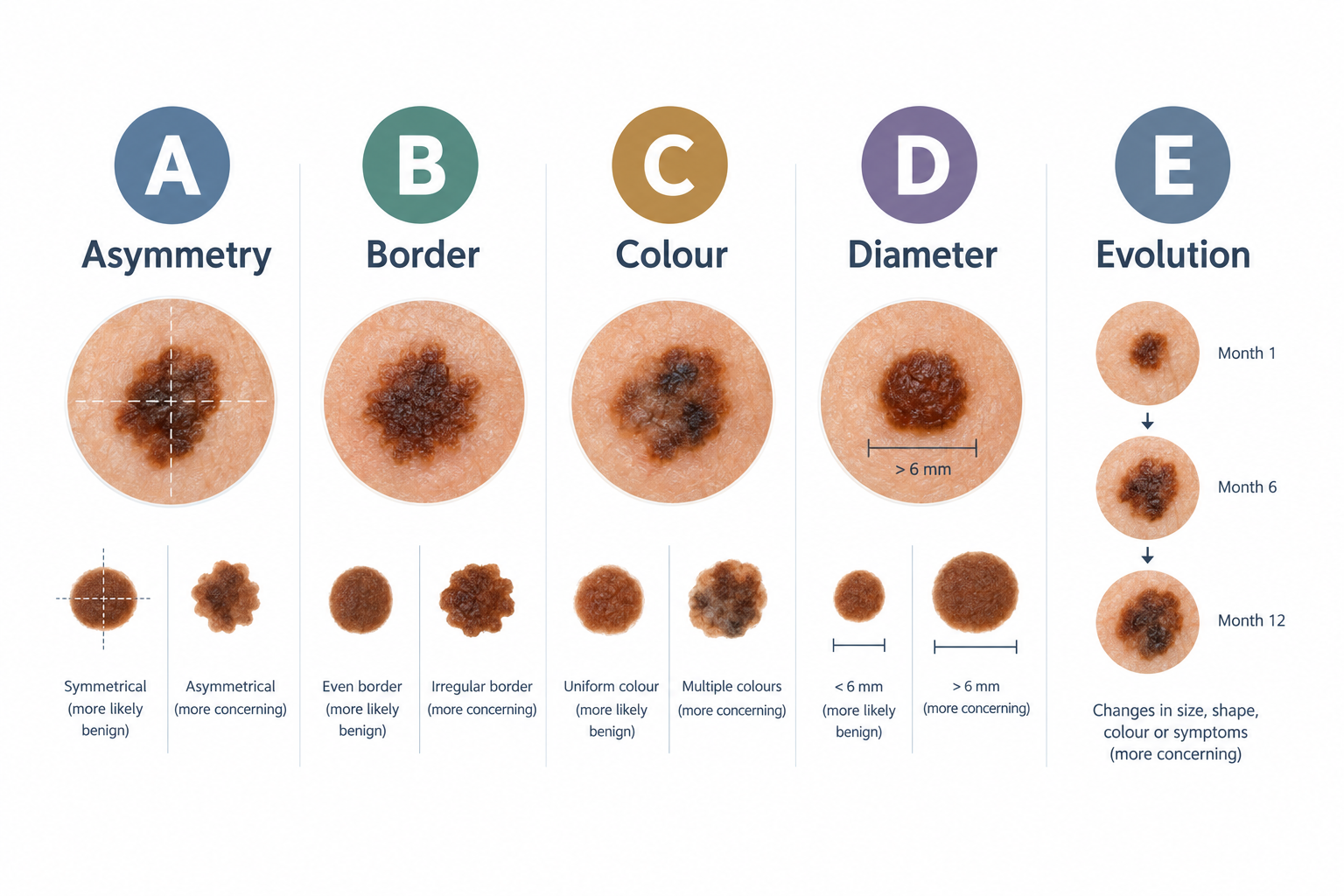
- Asymmetry: One half of the mole does not match the other in shape.
- Border: The edges are irregular, ragged, notched, or blurred rather than smooth and well-defined.
- Colour: The mole contains multiple shades — brown, black, red, white, or blue — or has changed colour noticeably.
- Diameter: The mole is larger than approximately 6 millimetres across, or has grown recently.
- Evolution: The mole has changed in size, shape, colour, or texture over weeks or months, or has developed new symptoms such as itching, bleeding, or crusting.
Any mole displaying one or more of these features should be assessed by a dermatologist or qualified clinician as a matter of priority, regardless of whether infection also appears to be present. Infection and malignant change are not mutually exclusive, and treating only the infection without investigating the mole itself could delay a critical diagnosis.
Additional symptoms that warrant urgent review include a mole that bleeds without obvious injury, a mole that ulcerates or develops an open sore that does not heal, a mole that has grown rapidly over a short period, or a mole that has become raised or nodular when it was previously flat.
Infected Mole vs Changing Mole: Understanding the Difference
Distinguishing between a mole that is infected and one that is undergoing potentially significant cellular change is not straightforward without clinical tools. Both can produce redness, swelling, and surface changes. However, there are some practical distinctions worth understanding.
An infected mole typically develops relatively quickly — often over one to three days — following a clear trigger such as trauma, shaving, or scratching. The redness tends to be concentrated around the mole rather than within it, and the mole’s underlying shape, colour, and border may remain unchanged once the infection resolves. Symptoms of infection often improve with appropriate treatment within a week to ten days.
A mole undergoing significant change, by contrast, tends to evolve more gradually over weeks or months, though some aggressive forms of skin cancer can progress more rapidly. The changes are typically within the mole itself — its colour, border, or surface texture — rather than in the surrounding skin alone. Importantly, a mole that appears infected but does not respond to standard infection treatment, or that continues to change after the infection has resolved, should be re-evaluated promptly.
How a Clinician Assesses a Changing or Infected Mole
When you present to a GP, dermatologist, or qualified skin specialist with a mole that has changed or appears infected, the clinician will typically begin with a detailed history. This includes how long the change has been present, whether there was any preceding trauma or irritation, whether the mole has changed in previous months or years, your personal history of sun exposure, and your family history of skin cancer or melanoma.
A physical examination of the mole will follow, often using a dermatoscope — a handheld illuminated magnification device that allows the clinician to examine the mole’s internal structure, colour patterns, and vascular features in detail that is not visible to the naked eye. Dermatoscopy significantly improves the accuracy of clinical assessment compared with visual inspection alone.
If infection is suspected, the clinician may take a swab to identify the causative organism and guide antibiotic treatment. If the mole’s appearance raises concern about malignant change, referral for excision and histological examination — where the removed tissue is analysed under a microscope — is the standard approach. Histology remains the definitive method for diagnosing or excluding skin cancer.
Treatment Options for an Infected Mole
Treatment depends on the underlying cause identified during clinical assessment. For a straightforward bacterial infection of the skin around a mole, a clinician may prescribe a course of topical or oral antibiotics, depending on the severity and extent of the infection. Keeping the area clean, avoiding further trauma, and refraining from picking or squeezing the mole are important supportive measures during treatment.
If the mole itself is the source of concern — either because of its appearance, its history of repeated infection, or clinical suspicion of malignant change — surgical excision is typically recommended. Excision involves removing the mole and a small margin of surrounding tissue under local anaesthetic. The removed tissue is then sent for histological analysis to confirm the diagnosis. This is both a treatment and a diagnostic procedure, and it is the most reliable way to assess a mole’s cellular composition.
Laser removal and other non-surgical mole treatments are generally not appropriate for moles that are clinically suspicious or that have shown signs of change, because these methods destroy the tissue rather than preserving it for histological examination. Any clinician offering laser removal of a mole should first carry out a thorough assessment to confirm the mole is benign.
What You Should Not Do at Home
It can be tempting to manage a sore or inflamed mole at home, particularly if the change seems minor. However, several common home approaches carry meaningful risks and should be avoided.
- Do not pick, squeeze, or scratch the mole. This can introduce bacteria, worsen existing infection, cause bleeding, and make clinical assessment more difficult.
- Do not apply over-the-counter wart or skin tag removal products. These products are not designed for use on moles and can cause chemical burns, scarring, and — critically — can destroy tissue that would otherwise be available for histological diagnosis.
- Do not attempt to remove the mole yourself. Home removal methods are unsafe, ineffective, and can delay diagnosis of a potentially serious condition.
- Do not assume that improvement in redness or swelling means the mole is safe. Infection can resolve while an underlying mole change continues to develop.
- Do not delay seeking assessment because you are concerned about what the clinician might find. Early detection of skin cancer, including melanoma, is strongly associated with better outcomes.
Risk Factors for Skin Cancer in Moles
Understanding which factors increase the likelihood that a changing mole may represent something more serious can help you make an informed decision about how urgently to seek assessment. The following factors are associated with a higher risk of melanoma or other skin cancers developing within or near a mole:
- A personal or family history of melanoma or other skin cancers
- A large number of moles — generally more than 50 — on the body
- The presence of atypical or dysplastic moles, which have irregular features but are not yet cancerous
- A history of significant or repeated sunburn, particularly in childhood or adolescence
- Prolonged cumulative sun exposure or use of sunbeds
- Fair skin, light hair, and light eyes, which are associated with reduced natural UV protection
- A weakened immune system, whether due to medical conditions or immunosuppressive medications
Having one or more of these risk factors does not mean that a changing mole is cancerous, but it does mean that prompt professional assessment is particularly important. People with multiple risk factors may benefit from regular skin surveillance appointments with a dermatologist.
When to See a Doctor: A Practical Guide
Many people are uncertain about when a mole change is serious enough to warrant a medical appointment. The following guidance may help, though it is not a substitute for individual clinical assessment.
Seek assessment within a few days if a mole has become red, swollen, painful, or is discharging fluid, particularly if there is no obvious cause such as recent trauma. While infection is a likely explanation, it is important to confirm this and to ensure the mole itself has not changed.
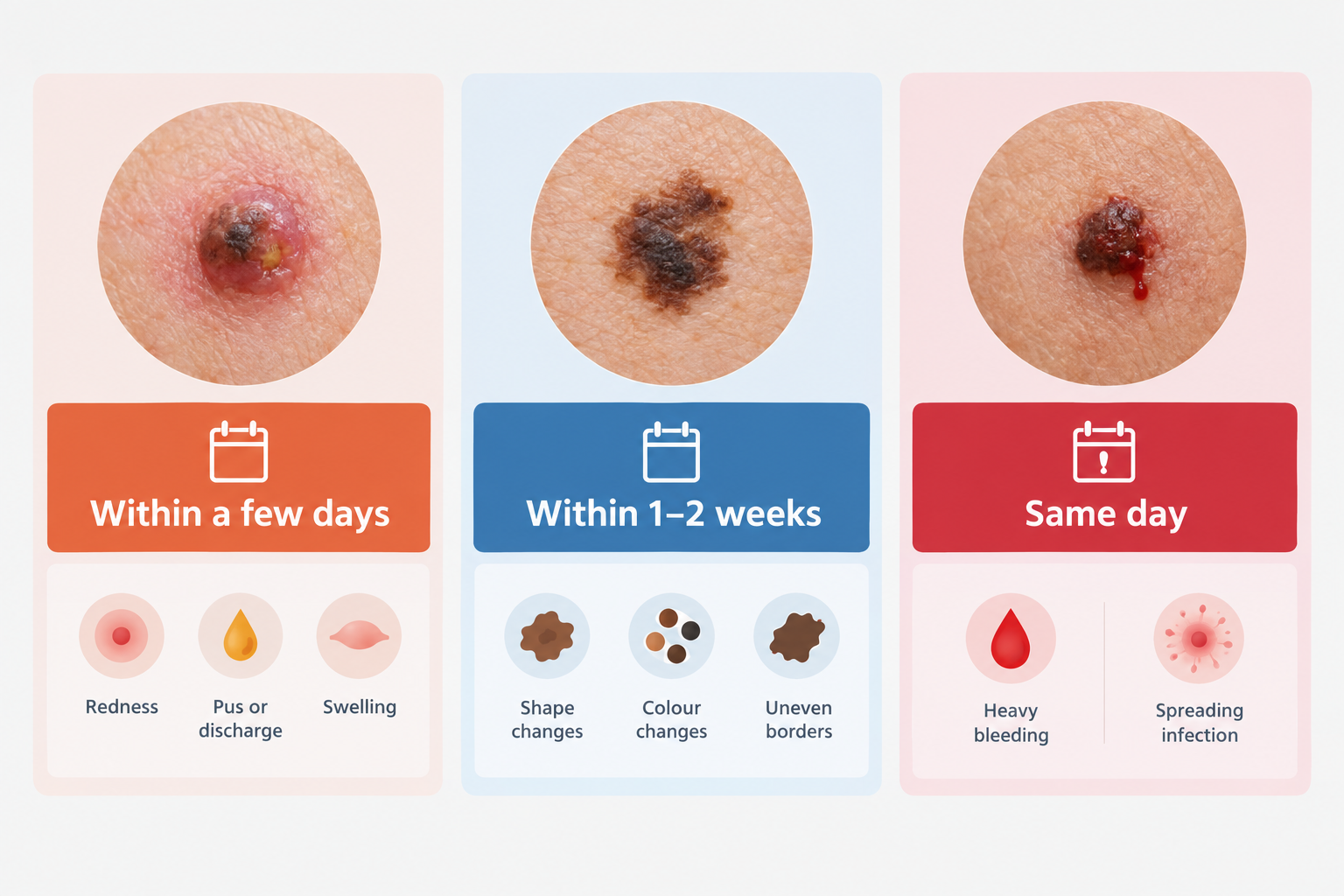
Seek urgent assessment — ideally within one to two weeks — if a mole has changed in size, shape, colour, or texture, if it has started to bleed without injury, if it has developed an irregular border, or if it has become raised or nodular. In the United Kingdom, the NHS two-week wait referral pathway exists specifically to ensure that potentially suspicious skin lesions are assessed by a specialist promptly.
Seek same-day or emergency assessment if a mole is bleeding heavily and the bleeding cannot be controlled, or if there are signs of rapidly spreading infection such as red streaking extending from the mole, fever, or significant swelling of the surrounding area.
If you are ever uncertain, erring on the side of seeking assessment is always the safer choice. A clinician can reassure you if the mole is benign and the infection is straightforward, and can act quickly if further investigation is needed.
Frequently Asked Questions
Can a mole become infected without any obvious injury?
Yes. Minor friction from clothing, repeated low-level irritation, or even a small scratch that goes unnoticed can create an entry point for bacteria. In some cases, folliculitis or a small cyst near the mole can cause inflammation without any clear preceding injury. If a mole appears infected without an obvious cause, clinical assessment is advisable to rule out other explanations for the change.
Is it normal for a mole to itch?
Occasional mild itching around a mole can result from dry skin, irritation, or minor inflammation. However, persistent itching within a mole — particularly if accompanied by other changes such as growth, colour change, or bleeding — is a symptom that should be assessed by a clinician. Itching alone is not a reliable indicator of malignancy, but it is a reason to seek professional review rather than to wait and observe.
How long does a mole infection take to heal?
A straightforward bacterial infection of the skin around a mole typically begins to improve within three to five days of appropriate antibiotic treatment and usually resolves within one to two weeks. If the area does not improve as expected, or if the mole itself continues to change after the infection has resolved, a follow-up appointment is important.
Can I use antiseptic cream on an infected mole?
Keeping the area clean is reasonable, and a mild antiseptic may help prevent the infection from worsening in the short term. However, antiseptic cream is not a substitute for clinical assessment and prescribed treatment where needed. It should not be used as a reason to delay seeking professional advice, particularly if the mole itself has changed or if the infection is not improving.
Will an infected mole leave a scar?
Minor infections that resolve without significant tissue damage are unlikely to cause permanent scarring. More severe infections, or those that have been picked or squeezed repeatedly, carry a greater risk of scarring. If the mole is subsequently excised for diagnostic or treatment purposes, a small linear scar is expected, though this typically fades over several months.
Should I photograph a changing mole?
Taking a clear photograph of a changing mole — ideally with a ruler or coin for scale — can be genuinely useful when you attend a clinical appointment. It allows the clinician to compare the mole’s current appearance with its appearance at an earlier date, which can help assess the rate and nature of any change. Smartphone photographs are generally adequate for this purpose.
When a Mole Changes: The Importance of Not Waiting
An infected mole is often a straightforward clinical problem with a clear treatment pathway, but the symptoms of infection and the warning signs of a more serious mole change can overlap in ways that make self-assessment unreliable. The most important practical step — whether a mole appears infected, has changed in appearance, or is simply behaving differently from before — is to seek assessment from a qualified clinician promptly.
Early evaluation allows infection to be treated effectively before it worsens, and ensures that any mole changes with a more serious underlying cause are identified at the earliest possible stage. Skin cancer, including melanoma, is among the most treatable cancers when detected early, and the outcomes associated with late diagnosis are considerably less favourable. No mole change is too minor to mention to a clinician, and no concern about a mole is too small to warrant a professional opinion.
If you have noticed any change in a mole — including redness, swelling, pain, bleeding, growth, or a shift in colour or border — arranging an assessment with your GP, a dermatologist, or a qualified skin specialist is the appropriate and responsible next step.