Mole Removal Healing Stages: A Week-by-Week Guide
After mole removal, most patients want to know exactly what their skin should look like at each stage of recovery. Healing progresses through predictable biological phases, but the timeline and appearance can vary depending on the removal method used, the size and depth of the mole, and individual skin characteristics. This guide explains what typically happens week by week, what is considered normal, and which signs may warrant a follow-up with your practitioner.
Key Takeaways
- Mole removal healing follows a sequence of inflammation, scabbing, re-epithelialisation, and scar remodelling that can span several weeks to months.
- The removal method — shave excision, surgical excision, or laser — significantly influences how the wound looks and how long full healing takes.
- Scabbing and mild redness in the first one to two weeks are normal; picking or scratching the scab increases the risk of scarring and infection.
- A pink or slightly raised mark is common for several weeks after the scab falls away and does not necessarily indicate a poor outcome.
- Any signs of spreading redness, increasing pain, discharge, or fever after the procedure should be assessed by a qualified practitioner promptly.
Why Healing Timelines Differ Between Patients
Mole removal healing is not a single uniform process. The depth of tissue removed, the technique used, and the location on the body all influence how quickly and cleanly the skin repairs itself. Shave excision, which removes a mole flush with the surrounding skin surface, typically produces a shallower wound that heals faster than a full surgical excision, which involves cutting deeper into the dermis and closing the wound with sutures. Laser removal is generally reserved for superficial, non-suspicious lesions and tends to produce a smaller wound footprint, though it is not appropriate for all mole types.
Individual factors also play a meaningful role. Skin that is naturally prone to hyperpigmentation may develop a darker post-inflammatory mark during healing. People with a tendency toward keloid or hypertrophic scarring may notice a thicker, raised scar forming over several weeks. Age, sun exposure history, smoking status, and immune function can all affect the pace and quality of wound healing. Understanding these variables helps set realistic expectations before the procedure begins.
The Four Biological Phases of Wound Healing
Regardless of the removal method, skin repair after mole removal follows four overlapping biological phases. Recognising these phases makes it easier to interpret what you see at each stage of recovery.
Phase 1: Haemostasis (Minutes to Hours)
Immediately after the procedure, the body works to stop any bleeding. Blood vessels constrict, platelets aggregate at the wound site, and a fibrin clot begins to form. This is the foundation on which subsequent healing is built. Patients may notice a small amount of bleeding or oozing directly after the procedure, which is normal and typically controlled with gentle pressure or a dressing applied by the practitioner.
Phase 2: Inflammation (Days 1–5)
The inflammatory phase begins within hours of the procedure and typically peaks over the first two to three days. The wound site may appear red, slightly swollen, and feel warm or tender to the touch. This is the immune system clearing debris and bacteria from the wound. A small amount of clear or pale yellow fluid seeping from the wound during this phase is generally normal wound exudate, not infection. True signs of infection — spreading redness, increasing warmth, thick yellow or green discharge, and fever — are distinct from normal inflammation and require prompt clinical assessment.
Phase 3: Proliferation and Re-epithelialisation (Days 5–21)
During the proliferative phase, new skin cells migrate across the wound surface, collagen is deposited, and new blood vessels form to support the healing tissue. This is when a scab typically forms and gradually shrinks as the skin underneath regenerates. The scab acts as a natural protective barrier and should be allowed to fall away on its own. Premature removal of the scab disrupts the fragile new skin beneath, increasing the risk of scarring and delaying healing. By the end of this phase, the wound surface is usually closed, though the underlying tissue continues to remodel.
Phase 4: Remodelling (Weeks 3 to 12 Months)
The remodelling phase is the longest and least visible stage of healing. Collagen fibres reorganise and mature, the scar gradually flattens, and redness or pink discolouration slowly fades. This phase can continue for up to 12 to 18 months in some cases, particularly for deeper excisions or in individuals prone to prolonged scar maturation. The final appearance of the scar is not fully established until remodelling is complete.
Week-by-Week Mole Removal Healing Stages
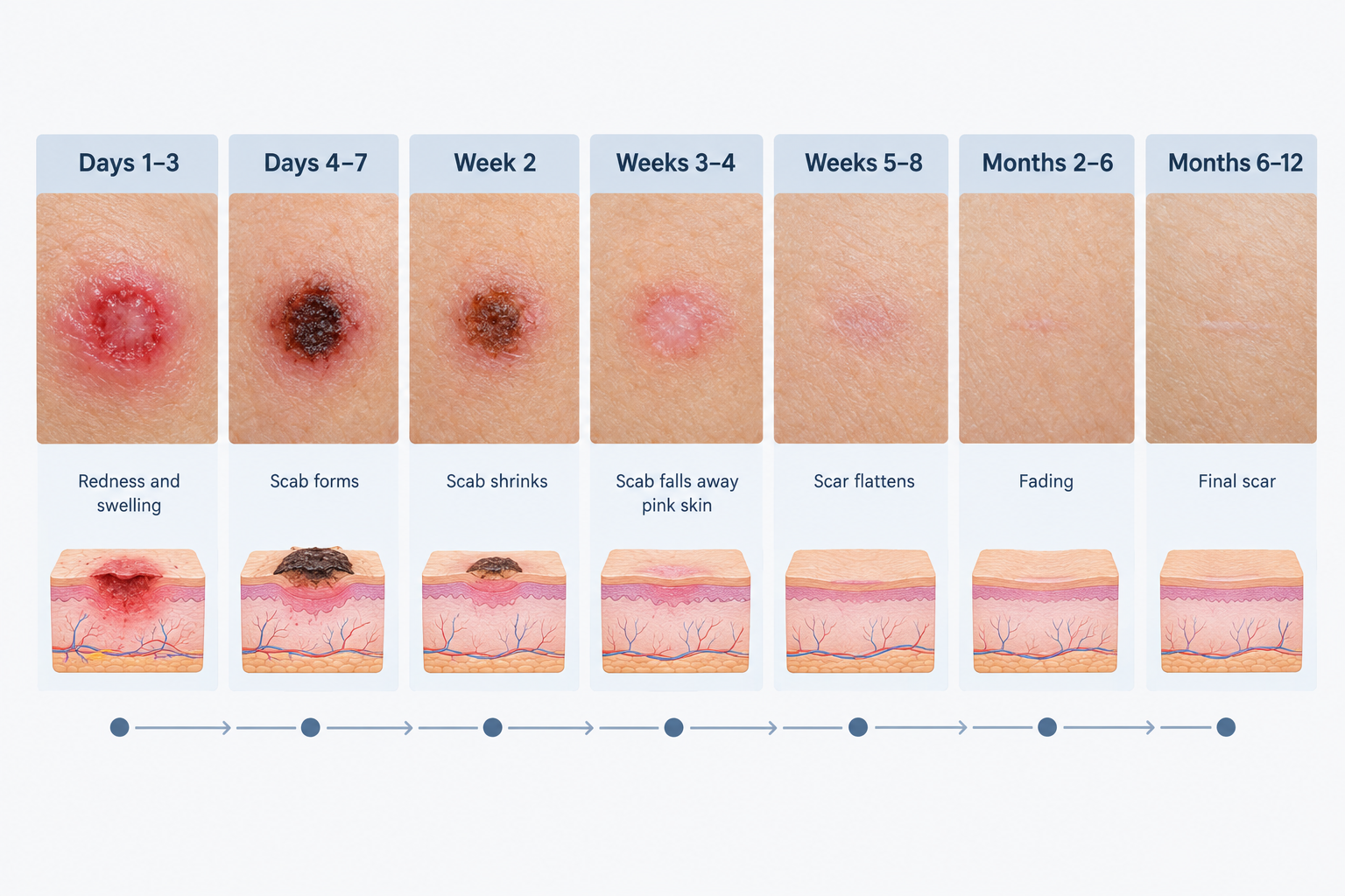
The following breakdown describes what patients typically observe at each stage after mole removal. These descriptions apply most directly to shave excision and surgical excision; laser removal may follow a slightly compressed timeline for superficial wounds.
Days 1–3: Immediate Post-Procedure Appearance
In the first 24 to 72 hours, the wound site is at its most visually dramatic. Redness, mild swelling, and tenderness are expected. A small dressing or adhesive bandage is usually applied immediately after the procedure to protect the wound and absorb any initial oozing. For surgical excisions with sutures, the stitches will be visible and the surrounding skin may be bruised. Patients are typically advised to keep the area clean and dry, avoid submerging the wound in water, and refrain from strenuous activity that could disrupt the wound edges.
Pain at this stage is usually mild to moderate and manageable with over-the-counter analgesics as recommended by the practitioner. Significant or worsening pain beyond the first day should be reported, as it may indicate a complication.
Days 4–7: Scab Formation
By the end of the first week, a scab will have formed over the wound in most cases. The scab may appear dark brown, reddish-brown, or black depending on the amount of dried blood and exudate present. This is a normal and protective part of the healing process. The surrounding skin may still appear slightly pink or red, and there may be mild itching as new skin cells proliferate beneath the scab surface.
For surgical excisions, sutures are often removed between five and ten days after the procedure, depending on the location and the practitioner’s assessment. Facial wounds typically heal faster and sutures may be removed earlier than those on the trunk or limbs. After suture removal, the wound edges may still appear slightly raised or pink.
Week 2: Scab Shrinkage and Early Skin Closure
During the second week, the scab typically begins to shrink and lift at the edges as new skin forms underneath. Itching may intensify slightly during this phase, which reflects active cellular regeneration. Moisturising the area with a gentle, fragrance-free product as advised by your practitioner can help manage discomfort without disrupting healing. The wound should not be scratched or picked, even if the scab appears loose.
By the end of week two, many shave excision wounds will have shed their scab entirely, revealing new pink skin beneath. Surgical excision sites may still retain part of the scab or show a visible linear scar line at this stage. The new skin is fragile and more susceptible to UV damage, making sun protection particularly important from this point onward.
Weeks 3–4: Scab Loss and Early Scar Formation
Most scabs will have fallen away naturally by the end of the third or fourth week. What remains is typically a flat or slightly raised pink or red mark. This post-inflammatory erythema — the persistent redness that follows wound closure — is a normal part of early scar formation and does not indicate a poor outcome. The mark may appear more noticeable in certain lighting conditions or after physical activity that increases blood flow to the skin.
At this stage, the new skin is still maturing. It may feel slightly different in texture compared to the surrounding skin and may be more sensitive to touch, temperature, or friction. Continued sun protection is essential, as UV exposure during early scar formation can trigger post-inflammatory hyperpigmentation, resulting in a darker mark that takes considerably longer to fade.
Weeks 5–8: Scar Maturation Begins
Between weeks five and eight, the scar typically begins to flatten and the redness starts to fade. For many patients with smaller shave excision wounds, the mark may become quite subtle during this period, blending progressively with the surrounding skin tone. For deeper surgical excisions, a linear scar line remains visible but should be softening in texture and lightening in colour.
Some patients notice that the scar appears slightly indented (atrophic) or slightly raised (hypertrophic) at this stage. A mildly raised scar that is still softening and fading is usually still within the normal range of healing. A scar that is becoming progressively thicker, harder, or extending beyond the original wound boundary may be developing into a hypertrophic or keloid scar, and this warrants a clinical review.
Months 2–6: Progressive Fading
Over the following months, collagen remodelling continues and the scar gradually matures. Pink or red discolouration typically fades to a lighter pink and then to a skin-toned or slightly paler mark. In individuals with darker skin tones, post-inflammatory hyperpigmentation — a darkening of the scar area — may develop or persist during this phase. This is caused by increased melanin production in response to inflammation and is generally temporary, though it can take six months or longer to resolve without targeted treatment.
Scar massage, silicone gel or sheeting, and consistent broad-spectrum sun protection are commonly recommended during this phase to support optimal scar maturation. These measures are most effective when started after the wound has fully closed and the practitioner has confirmed the wound is healing appropriately.
Months 6–12 and Beyond: Final Scar Appearance
The final appearance of a mole removal scar is generally not established until at least six to twelve months after the procedure, and in some cases longer. By this point, most scars will have faded significantly and flattened considerably. A small, pale, flat mark is a common outcome for straightforward shave excisions. Surgical excision scars typically leave a fine linear mark whose visibility depends on the original wound size, location, closure technique, and individual healing response.
If the scar remains raised, thickened, itchy, or discoloured beyond twelve months, a range of scar treatment options may be worth discussing with a dermatologist or aesthetic practitioner. These can include topical treatments, steroid injections, laser resurfacing, or microneedling, depending on the scar type and severity.
What Normal Healing Looks Like vs. Warning Signs
Understanding the difference between expected healing changes and signs that require clinical attention is important for patient safety.
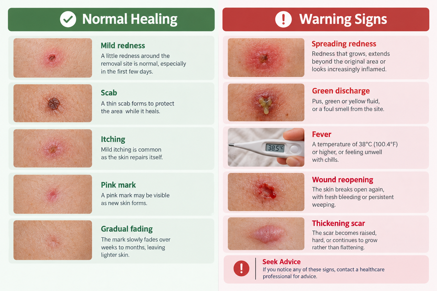
Normal Healing Signs
- Mild redness, swelling, and tenderness in the first three to five days
- Scab formation over the wound surface during the first week
- Itching as new skin forms beneath the scab
- Pink or red discolouration of the healing mark after the scab falls away
- Gradual flattening and fading of the scar over weeks and months
- Slight texture difference between the scar and surrounding skin during early maturation
Signs That Warrant Prompt Clinical Assessment
- Spreading redness or red streaking extending beyond the wound edges
- Increasing pain or throbbing after the first few days
- Thick yellow or green discharge from the wound
- Fever or feeling generally unwell after the procedure
- Wound edges separating or the wound reopening
- A scar that continues to grow thicker, harder, or larger beyond the wound boundary
- Any new or changing pigmentation within or around the treated area that was not discussed with your practitioner
Factors That Can Affect Healing and Scar Outcome
Several modifiable and non-modifiable factors influence how well the skin heals after mole removal. Being aware of these can help patients take practical steps to support recovery.
Sun Exposure
UV radiation is one of the most significant factors affecting scar appearance. Exposing a healing wound or immature scar to sunlight can cause post-inflammatory hyperpigmentation, making the mark darker and more persistent. Broad-spectrum SPF 30 or higher sunscreen should be applied to the area daily once the wound has closed, and physical protection such as clothing or a dressing is advisable during the early healing weeks.
Wound Care Adherence
Following the aftercare instructions provided by your practitioner — including keeping the wound clean, applying any recommended ointments, and attending follow-up appointments — directly affects healing quality. Neglecting wound care increases the risk of infection and suboptimal scarring.
Smoking
Smoking impairs wound healing by reducing oxygen delivery to healing tissue and impairing immune function. Patients who smoke may experience slower healing and a higher risk of wound complications. Stopping smoking before and after the procedure, where possible, is generally advisable.
Skin Type and Scar Tendency
Individuals with Fitzpatrick skin types IV to VI are at higher risk of post-inflammatory hyperpigmentation. Those with a personal or family history of keloid scarring are at elevated risk of developing a raised, thickened scar that extends beyond the original wound. This risk should be discussed with the practitioner before the procedure, as it may influence the choice of removal method and aftercare approach.
Location on the Body
Wounds on areas subject to movement or tension — such as the chest, shoulders, or joints — are more prone to widened or hypertrophic scars than wounds on the face, where the skin is less mobile and blood supply is rich. Facial wounds generally heal faster and with less visible scarring than wounds on the trunk or limbs.
Scar Treatment Options If Healing Is Unsatisfactory
If the scar remains more visible than expected after full maturation, several evidence-supported options may be worth discussing with a qualified practitioner. Silicone gel and silicone sheeting are among the most widely recommended non-invasive approaches for hypertrophic scars and are most effective when used consistently over several months. Intralesional corticosteroid injections can help flatten raised or thickened scars by reducing collagen overproduction. Laser treatments, including fractional laser resurfacing and pulsed dye laser, may improve scar texture, redness, and overall appearance, though multiple sessions are typically required and results vary. Microneedling is sometimes used to improve atrophic or textured scars by stimulating collagen remodelling. The most appropriate option depends on the scar type, skin tone, and individual circumstances, and a clinical assessment is necessary before starting any scar treatment.
Frequently Asked Questions
How long does it take for a mole removal wound to fully heal?
Surface wound closure typically occurs within two to four weeks depending on the removal method and wound size. However, scar remodelling continues for up to twelve months or longer, and the final scar appearance is not established until this process is complete.
Is it normal for the area to look worse before it looks better?
Yes. The wound site often appears most noticeable during the first one to two weeks when the scab is present and surrounding redness is at its peak. Once the scab falls away and inflammation subsides, the appearance typically improves progressively over the following weeks and months.
What should I do if my scab falls off early?
If the scab detaches before the wound has fully closed underneath, keep the area clean and moist with a gentle ointment as advised by your practitioner, and protect it from sun exposure and friction. Contact your practitioner if the wound appears open, is bleeding, or shows signs of infection.
Can I wear makeup over the healing area?
Most practitioners advise avoiding makeup directly over an open wound or fresh scab. Once the wound has fully closed and the scab has naturally fallen away, mineral-based or non-comedogenic makeup is generally considered lower risk, but it is best to confirm this with your practitioner based on your specific wound.
Will the scar be permanent?
Some degree of permanent mark is possible after mole removal, particularly for deeper surgical excisions. However, many scars fade significantly over twelve months and become difficult to notice. The final outcome depends on the removal method, wound size, location, individual healing response, and aftercare adherence.
Why is my scar itchy weeks after the procedure?
Itching during scar maturation is common and reflects ongoing collagen remodelling and nerve regeneration within the healing tissue. It is generally not a sign of infection. Gentle moisturising and silicone gel application can help manage itching. If itching is severe or accompanied by other symptoms, a clinical review is advisable.
When should I be concerned about changes in the treated area?
Any new or changing pigmentation, irregular borders, or unusual growth within or around the treated area should be assessed by a qualified practitioner, particularly if the original mole was removed for diagnostic reasons. Routine follow-up appointments allow the practitioner to monitor healing and identify any concerns early.
What to Expect from Your Mole Removal Recovery
Mole removal healing is a gradual process that unfolds over weeks and months rather than days. The most visually active phase — scabbing, redness, and early scar formation — typically resolves within the first four to six weeks, but the underlying tissue continues to remodel long after the surface appears healed. Understanding each stage of recovery helps patients distinguish normal healing from signs that need attention, and supports better aftercare decisions throughout the process.
Consistent sun protection, careful wound care, and avoiding habits such as picking or scratching the healing area are among the most practical steps patients can take to support a good outcome. If the scar remains prominent after twelve months, or if healing appears to be progressing abnormally at any stage, a follow-up consultation with a dermatologist or qualified aesthetic practitioner can help identify whether any additional treatment is appropriate. Every patient’s healing journey is individual, and a personalised assessment remains the most reliable guide to managing recovery effectively.