Cancerous Pink Mole Signs: What to Look For and When to Act
A pink mole can be entirely harmless, but certain characteristics make it a potential early warning sign of skin cancer — including some of the most dangerous types. Because pink or flesh-coloured lesions lack the dark pigment most people associate with melanoma, they are frequently dismissed or misidentified, which can lead to delayed diagnosis and a more advanced stage at detection. Understanding what distinguishes a benign pink mole from a cancerous one could make a meaningful difference to your outcome.
Key Takeaways
- Not all cancerous moles are dark brown or black — amelanotic melanoma, basal cell carcinoma, and squamous cell carcinoma can all appear pink, flesh-coloured, or pearly.
- The ABCDE rule (Asymmetry, Border, Colour, Diameter, Evolving) applies to pink moles and remains one of the most reliable self-assessment tools available to patients.
- Multiple shades of pink within a single lesion is a specific dermoscopic marker associated with early amelanotic melanoma and should prompt professional evaluation.
- Amelanotic melanoma carries a lower five-year survival rate than pigmented melanoma largely because delayed diagnosis allows the cancer to reach a more advanced stage before treatment begins.
- A full-body skin exam by a qualified dermatologist, combined with regular skin self-exam, is the most effective strategy for catching suspicious pink moles early.
What Is a Pink Mole?
Most moles contain melanin, the pigment produced by melanocytes (pigment-producing skin cells), which gives them their characteristic brown or black colour. A pink mole — sometimes described as flesh-coloured, salmon-coloured, or skin-toned — contains little or no melanin. This reduced pigmentation can occur in both benign and malignant lesions, which is precisely what makes pink moles clinically significant. Benign pink moles are common and include ordinary compound nevi that have lost pigment over time, as well as dysplastic nevi (atypical moles) that were never deeply pigmented. However, the same pale appearance is a hallmark of several types of skin cancer, making visual assessment alone unreliable without professional evaluation.
Types of Skin Cancer That Can Appear Pink
Several distinct skin cancers present as pink, pearly, or flesh-coloured lesions. Recognising which type may be involved helps explain why the warning signs matter and why prompt assessment is important.
Amelanotic Melanoma
Amelanotic melanoma is a subtype of melanoma that produces little or no visible pigment. It accounts for roughly 2–8% of all melanomas and is widely considered the most dangerous pink skin lesion because it so closely mimics benign conditions such as a pimple, scar, eczema patch, or pyogenic granuloma. Because it lacks the dark colouring that most people associate with melanoma warning signs, amelanotic melanoma is frequently overlooked during both self-examination and initial clinical assessment. Research published in peer-reviewed dermatology literature highlights that the presence of multiple shades of pink within a single lesion — visible under dermoscopy — is a meaningful early indicator of amelanotic melanoma, a nuance rarely communicated in standard patient-facing resources. Nodular melanoma, a fast-growing subtype, can also present with minimal pigmentation and a pink or red raised appearance.
Basal Cell Carcinoma
Basal cell carcinoma (BCC) is the most common form of skin cancer and frequently appears as a pearly bump or shiny pink nodule, sometimes with visible blood vessels (telangiectasia) on its surface. BCC grows slowly and rarely spreads to other parts of the body, but it can cause significant local tissue destruction if left untreated. It most commonly develops on sun-exposed areas such as the face, ears, neck, and scalp. A non-healing sore that repeatedly crusts, bleeds, and reforms is a classic presentation of BCC and should always be assessed by a dermatologist.
Squamous Cell Carcinoma
Squamous cell carcinoma (SCC) typically presents as a firm, pink, scaly patch or raised nodule that may ulcerate or bleed. SCC carries a higher risk of spreading to lymph nodes than BCC, particularly when it arises on the lips, ears, or in immunosuppressed individuals. Actinic keratosis — a rough, scaly precancerous lesion caused by cumulative UV radiation — can appear pink or red and is considered a precursor to SCC. Treating actinic keratoses early reduces the risk of progression to invasive cancer.
Merkel Cell Carcinoma
Merkel cell carcinoma is a rare but aggressive skin cancer that often appears as a fast-growing, painless, flesh-coloured or pink nodule, most commonly on the face, head, or neck. It is more likely to spread than either BCC or SCC and requires prompt specialist assessment. Although uncommon, its pink appearance means it can be confused with a benign cyst or lymph node enlargement.
Warning Signs of a Cancerous Pink Mole
The challenge with pink moles is that their lack of pigment removes one of the most recognisable cancer warning signs. The following features, individually or in combination, should prompt a professional evaluation rather than a wait-and-see approach.
The ABCDE Rule Applied to Pink Moles
The ABCDE rule was developed to help both patients and clinicians identify potentially malignant moles. It applies equally to pink and pigmented lesions, though some criteria require slight reinterpretation for non-pigmented moles.
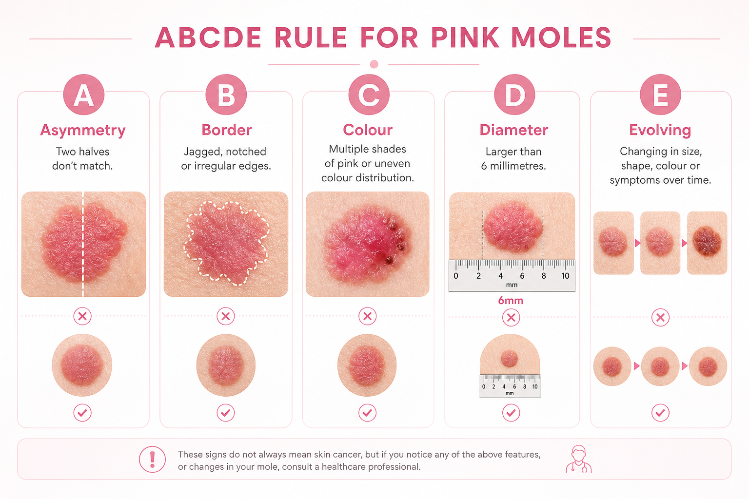
- Asymmetry: One half of the mole does not match the other. Benign moles tend to be symmetrical; cancerous lesions are often irregular in shape.
- Border: An irregular border — jagged, notched, blurred, or poorly defined edges — is a warning sign. Benign moles typically have smooth, well-defined margins.
- Colour: For pink moles, look for multiple shades of pink, red, or white within the same lesion. Variation in colour across a single mole is more concerning than a uniform tone, even if that tone is pale.
- Diameter: Lesions larger than 6 millimetres (roughly the size of a pencil eraser) warrant attention, though melanomas can be smaller, particularly at an early stage.
- Evolving: Any mole that is changing in size, shape, colour, or texture — or that begins to itch, bleed, crust, or ooze — should be assessed promptly. An evolving mole is one of the most clinically significant warning signs regardless of colour.
The Ugly Duckling Sign
The ugly duckling sign is a complementary self-assessment concept: a mole that looks noticeably different from the others on your body — the one that stands out — deserves closer attention. Most benign moles on a given person share a similar general appearance. A pink mole that looks distinctly unlike your other moles, particularly if it is growing or changing, should be evaluated by a dermatologist even if it does not clearly meet ABCDE criteria.
Additional Features That Raise Concern
- A raised, dome-shaped, or nodular surface on a previously flat lesion
- Ulceration or a sore that will not heal despite several weeks passing
- Crusting, oozing, or bleeding without injury
- Satellite lesions — small new spots appearing near an existing mole
- Spread of colour or texture into the surrounding skin
- A pearly or waxy surface with visible blood vessels
Normal Pink Mole vs. Cancerous Pink Mole: A Comparison
| Feature | Benign Pink Mole | Potentially Cancerous Pink Mole |
|---|---|---|
| Shape | Symmetrical, round or oval | Asymmetrical, irregular |
| Border | Smooth, well-defined | Irregular, jagged, or blurred |
| Colour | Uniform single shade of pink | Multiple shades of pink, red, or white |
| Size | Usually under 6mm, stable | Often over 6mm or actively growing |
| Surface | Smooth or slightly raised, intact | Raised, ulcerated, crusting, or bleeding |
| Change over time | Stable for months or years | Changing in size, shape, or texture |
| Healing | Does not bleed or crust spontaneously | May bleed, crust, or fail to heal |
Pink Moles on Different Skin Tones
Most published guidance on mole warning signs is written with fair-skinned patients in mind, but skin cancer — including amelanotic melanoma — occurs across all skin tones. On medium and darker skin, a pink or flesh-coloured lesion may appear as a slightly lighter patch relative to the surrounding skin, or it may present with a reddish or translucent quality rather than an obvious pink hue. The ABCDE rule remains applicable regardless of skin tone, but the colour criterion requires contextual interpretation: look for any unusual variation in tone within the lesion rather than expecting a specific shade of pink. People with darker skin tones are also more likely to develop melanoma in less sun-exposed locations — such as the palms, soles, under the nails, or on mucous membranes — where lesions may go unnoticed for longer. This contributes to later-stage diagnosis in some populations and underscores the importance of full-body skin checks that include these areas.
Pink Moles in Hard-to-See Locations
Cancerous pink moles can develop anywhere on the body, including areas that are difficult to examine during a routine self-check. The scalp, the skin between the toes, the soles of the feet, the genital region, and the area beneath the nails are all sites where melanoma and other skin cancers have been documented. These locations are frequently missed during self-examination and may not be routinely inspected during a general medical appointment. A full-body skin exam performed by a trained dermatologist specifically includes these harder-to-reach areas and is the most reliable way to ensure that lesions in concealed locations are not overlooked.
Conditions That Can Mimic a Cancerous Pink Mole
One of the most clinically significant challenges with amelanotic melanoma is how convincingly it can resemble benign conditions. Patients and even clinicians have mistaken amelanotic melanoma for a pimple, an inflamed cyst, a healing scar, a patch of eczema, or a stretch mark. Several benign skin conditions share a pink or flesh-coloured appearance:
- Pyogenic granuloma: A rapidly growing, bright red or pink vascular lesion that bleeds easily. It is benign but can look alarming and may resemble nodular melanoma.
- Dermatofibroma: A firm, slightly raised pink or skin-coloured nodule, usually on the legs, that dimples inward when pinched.
- Sebaceous hyperplasia: Enlarged sebaceous glands that appear as small, yellowish-pink bumps, most often on the face.
- Molluscum contagiosum: Viral skin lesions that appear as small, pearly pink or flesh-coloured bumps with a central dimple.
- Inflamed seborrhoeic keratosis: A common benign growth that can become irritated and appear pink or red.
Because these conditions can look similar to early skin cancer, any new or changing pink lesion that does not resolve within a few weeks should be assessed professionally rather than assumed to be benign.
Risk Factors for Cancerous Pink Moles
Certain factors increase the likelihood that a pink mole may be or become malignant. Understanding your personal risk profile helps determine how frequently you should undergo skin cancer screening and how vigilantly you should monitor your skin.
- UV radiation exposure: Cumulative sun exposure and the use of tanning beds are the most significant modifiable risk factors for all types of skin cancer. UV radiation damages the DNA in skin cells, increasing the likelihood of malignant change.
- Fair skin, light hair, and light-coloured eyes: People with less melanin in their skin have reduced natural protection against UV damage.
- Family history of skin cancer: A first-degree relative with melanoma increases personal risk, particularly if the family history includes multiple cases or early-onset disease.
- Personal history of skin cancer: Having had one melanoma or non-melanoma skin cancer significantly raises the risk of developing another.
- Large number of moles: Having 50 or more moles, or a history of dysplastic nevi (atypical moles), is associated with elevated melanoma risk.
- Blistering sunburns: Particularly those occurring in childhood or adolescence, blistering sunburns are associated with a meaningfully higher lifetime risk of melanoma.
- Immunosuppression: Organ transplant recipients and others on long-term immunosuppressive therapy have a substantially higher risk of developing skin cancers, including SCC.
How Pink Moles Are Diagnosed
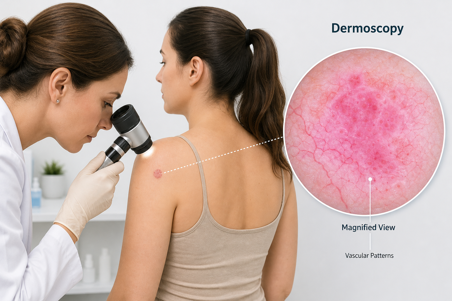
When a dermatologist examines a suspicious pink mole, the assessment typically begins with a visual inspection followed by dermoscopy — a technique that uses a handheld device with magnification and polarised light to examine the surface structures of a lesion in detail. Dermoscopy significantly improves diagnostic accuracy compared with the naked eye alone and allows clinicians to identify subtle features such as vascular patterns, multiple shades of pink, and structural irregularities that are not visible without magnification. In some specialist centres, reflectance confocal microscopy — a non-invasive imaging technique that provides near-histological detail of skin layers — may be used to further characterise ambiguous lesions before a decision about biopsy is made.
If a lesion is considered suspicious after clinical and dermoscopic assessment, a skin biopsy is performed. A small sample of the lesion is removed and examined by a dermatopathologist under a microscope. The biopsy result determines whether the lesion is benign, dysplastic, or malignant, and if malignant, the Breslow depth (the thickness of the tumour) helps guide staging and treatment planning. Emerging AI-based image analysis tools are beginning to assist in the triage of suspicious lesions, though these technologies are not yet a replacement for clinical assessment and biopsy.
Treatment Options for Cancerous Pink Moles
Treatment depends on the type of skin cancer identified, its stage, location, and the individual patient’s health. For most early-stage melanomas and non-melanoma skin cancers, surgical excision — removing the lesion along with a margin of surrounding healthy tissue — is the primary treatment. The width of the excision margin is determined by the tumour type and Breslow depth. For basal cell carcinoma, particularly on the face, Mohs surgery (a precise layer-by-layer excision technique) is frequently used to maximise tissue preservation while ensuring complete removal.
Advanced or metastatic melanoma may require additional treatment beyond surgery. Immunotherapy — which uses the body’s immune system to target cancer cells — and targeted therapies such as BRAF inhibitors and MEK inhibitors have significantly improved outcomes for patients with advanced melanoma over the past decade. Radiation therapy may be used in specific circumstances, such as when surgery is not feasible or when treating lymph node involvement.
Prognosis: Why Early Detection Matters
The five-year survival rate for melanoma detected at an early, localised stage is approximately 98–99%. When melanoma has spread to regional lymph nodes, the five-year survival rate falls to around 68%, and for distant metastatic disease it drops further still. Amelanotic melanoma is associated with a lower overall five-year survival rate — approximately 88% compared with around 95% for pigmented melanoma — and the primary reason is delayed diagnosis. Because amelanotic melanoma lacks the dark pigment that triggers concern, patients and clinicians are less likely to identify it early, meaning it is more often detected at a later stage when treatment is more complex and outcomes are less favourable. This survival gap is not an inherent property of the cancer’s biology alone; it is substantially driven by the time lost before diagnosis. Early detection through regular skin self-exam and professional skin cancer screening directly addresses this risk.
How to Monitor Your Moles at Home
A monthly skin self-exam is a practical and evidence-supported habit for anyone with moles, particularly those with known risk factors. Perform the exam in good lighting, using a full-length mirror and a hand mirror to check areas that are difficult to see directly. Systematically examine the scalp (use a comb or hair dryer to part the hair), face, neck, chest, abdomen, arms, hands, back, buttocks, legs, feet, and the skin between the toes. Ask a partner to help check the back and scalp if needed.
Photographing your moles using a smartphone provides a useful baseline for comparison over time. Several smartphone applications are designed specifically for mole tracking, allowing you to document the size, shape, and colour of individual lesions and compare them across months. While these apps are not a substitute for professional assessment, they can help you notice changes that might otherwise be missed between annual dermatologist appointments. Any mole that appears new, grows, changes colour, or develops any of the ABCDE warning signs should be assessed by a dermatologist promptly rather than waiting for a scheduled review.
Pink Moles in Children and Teenagers
New moles appearing in childhood and adolescence are common and usually benign. However, parents should be aware that congenital melanocytic nevi — moles present at birth or appearing in the first few months of life — carry a slightly elevated lifetime risk of malignant transformation, particularly when they are large. Any mole in a child or teenager that grows rapidly, changes shape, bleeds, or looks distinctly different from other moles on the body should be evaluated by a paediatric dermatologist. Melanoma in children is rare but does occur, and the same ABCDE warning signs apply regardless of age.
Frequently Asked Questions
Can a normal mole be pink?
Yes. Many benign moles are pink or flesh-coloured, particularly in people with fair skin or in moles that have lost pigment over time. A pink mole is not automatically a cause for alarm, but any pink mole that is new, growing, changing, or has irregular features should be assessed by a dermatologist.
What does amelanotic melanoma look like?
Amelanotic melanoma typically appears as a pink, red, or flesh-coloured lesion that may be flat or slightly raised. It often lacks the dark pigmentation associated with typical melanoma and can resemble a pimple, scar, or benign skin growth. Under dermoscopy, the presence of multiple shades of pink and atypical vascular patterns are important diagnostic clues.
Is a pink pearly bump always basal cell carcinoma?
Not necessarily, but a pearly bump — particularly one that is shiny, has visible blood vessels, and appears on a sun-exposed area — is a classic presentation of basal cell carcinoma and should be evaluated by a dermatologist. Other conditions, including benign cysts and sebaceous hyperplasia, can also appear pearly or pink.
How quickly should I see a doctor about a pink mole?
If a pink mole is new, growing, changing, bleeding, crusting, or has any ABCDE warning signs, seek a dermatologist appointment within a few weeks rather than waiting for a routine annual check. If a lesion is bleeding heavily or changing very rapidly, seek assessment sooner. Early evaluation is always preferable to delayed diagnosis.
Can sunscreen prevent cancerous pink moles?
Broad-spectrum sunscreen with a high SPF, combined with protective clothing and avoidance of tanning beds, reduces cumulative UV radiation exposure and lowers the overall risk of skin cancer. Sunscreen does not eliminate risk entirely, and it does not treat existing lesions, but consistent sun protection is one of the most evidence-supported strategies for reducing skin cancer risk over a lifetime.
Are pink moles more dangerous than dark moles?
Pink moles are not inherently more dangerous, but they are more likely to be overlooked because they do not match the typical image of a cancerous mole. This increases the risk of delayed diagnosis, which in turn affects treatment outcomes. Any mole — regardless of colour — that displays warning signs warrants professional evaluation.
When to See a Dermatologist About a Pink Mole
A pink mole that is stable, symmetrical, has a smooth border, a uniform colour, and has not changed in months or years is unlikely to require urgent attention, though it is reasonable to mention it at your next routine appointment. However, professional evaluation is warranted without delay if a pink mole is new and growing, has an irregular border or asymmetrical shape, contains multiple shades of pink or red, is larger than 6 millimetres, is raised or nodular, bleeds or crusts without injury, or simply looks different from your other moles. Annual skin cancer screening with a dermatologist — including a full-body skin exam — is recommended for anyone with significant risk factors, including a family history of skin cancer, a personal history of skin cancer, fair skin, or a large number of atypical moles. For those without specific risk factors, discussing the appropriate frequency of professional skin checks with a GP or dermatologist is a sensible starting point. The goal is not to create unnecessary anxiety about every pink spot, but to ensure that genuinely suspicious lesions are identified and assessed before they have the opportunity to progress.