Can Removing a Mole Cause Cancer? What the Evidence Actually Shows
The fear that removing a mole might trigger or spread cancer is one of the most common concerns patients raise before a skin procedure — and it is entirely understandable. The short answer, supported by dermatological evidence, is that professionally performed mole removal does not cause cancer. However, the fuller picture involves important nuances around incomplete removal, the risks of DIY approaches, and what happens when a mole that was already cancerous is removed. This article explains the biology behind that reassurance, addresses the real risks that do exist, and helps you understand when and how to seek appropriate care.
Key Takeaways
- Professional mole removal performed by a trained dermatologist or skin specialist does not cause cancer — a scalpel cannot trigger the DNA mutations that lead to malignancy.
- The greatest real risk is incomplete removal, which can leave residual cells behind and delay diagnosis if the mole contained abnormal or cancerous tissue.
- DIY mole removal is unsafe: it carries risks of infection, scarring, and — most critically — missed or delayed diagnosis of a potentially cancerous lesion.
- After any mole removal, the tissue should be sent for pathological analysis to confirm whether the cells were benign, precancerous, or malignant.
- Mole removal does not eliminate your ongoing skin cancer risk; regular skin self-examination and annual dermatologist checks remain important regardless of previous removals.
Why People Fear That Mole Removal Causes Cancer
This concern has circulated for generations, often passed down as informal advice: “Don’t touch a mole or you’ll disturb it and cause cancer.” The fear is understandable because cancer is sometimes diagnosed shortly after a mole is removed — leading people to assume the removal was the cause. In reality, the cancer was almost always pre-existing; the removal either revealed it through pathological analysis or, in cases of incomplete removal, left residual cancerous cells that continued to grow. The removal itself was not the cause.
A second source of anxiety is the idea that physically disrupting a mole — cutting into it — might cause cancerous cells to spread through the bloodstream or lymphatic system. This concern is not supported by clinical evidence for standard mole removal. Cancer requires specific genetic mutations in cells; a surgical instrument cannot induce those mutations. What a poorly performed or incomplete removal can do is leave abnormal cells behind, which is a meaningfully different problem and one that professional technique is specifically designed to prevent.
How Cancer Actually Develops: The Biology in Plain Language
Understanding why mole removal does not cause cancer requires a brief look at how cancer actually begins. Skin cancers, including malignant melanoma, develop when melanocytes — the pigment-producing cells in the skin — accumulate DNA mutations, typically driven by ultraviolet radiation from sun or UV exposure. These mutations cause cells to grow and divide uncontrollably. This is a biological process that unfolds over months or years at the cellular level; it is not something that can be initiated by a scalpel making contact with skin.
A benign melanocytic nevus (a common mole) is simply a cluster of melanocytes that have grown in a controlled, non-invasive way. Removing that cluster surgically does not alter the DNA of surrounding cells or create new mutations. The surrounding skin cells remain exactly as they were before the procedure. What removal does do — when performed correctly — is eliminate the mole tissue entirely, which is why professional mole removal is considered a safe and routine procedure when carried out by a qualified practitioner.
The Real Risks: What Can Actually Go Wrong
While mole removal does not cause cancer, there are genuine risks associated with how and by whom a mole is removed. Understanding these risks helps clarify why professional assessment matters.
Incomplete Removal and Residual Cells
Incomplete removal is the most clinically significant risk associated with mole removal procedures. If a mole is not excised with adequate clear surgical margins — meaning a small border of healthy tissue around the mole — some cells may be left behind. For a benign mole, this typically results in mole recurrence or regrowth, which is cosmetically inconvenient but not dangerous. For a mole containing precancerous changes or malignant cells, residual tissue left in place can continue to develop, potentially progressing or delaying the diagnosis of a more serious condition.
Recurrence rates vary meaningfully by removal method. Shave excision, which removes the mole at or just below the skin surface, carries a higher recurrence rate — estimated in some studies at between 11% and 33% for dysplastic nevi — because it does not always reach the deepest layer of the mole. Surgical excision, which removes the full thickness of the mole along with a margin of surrounding tissue and is closed with sutures, carries a recurrence rate of well under 1% when performed with adequate margins. This difference is one reason why the choice of removal method matters, particularly for atypical or suspicious lesions.
Pseudomelanoma: A Nuanced Complication
A less widely known phenomenon called pseudomelanoma can occur after incomplete shave removal of a benign mole. When a mole regrows after partial removal, the regenerating tissue can display cellular features — under microscopic examination — that resemble melanoma, even though the lesion is actually benign. This can cause diagnostic confusion and unnecessary anxiety or further procedures. A pathologist experienced in skin pathology can usually distinguish pseudomelanoma from true malignancy, but the situation underscores why complete, properly performed removal and accurate pathological analysis are important from the outset.
What Happens When a Removed Mole Was Already Cancerous
If a mole that appeared benign turns out to contain malignant melanoma or other cancerous cells on pathological examination, the removal itself did not cause the cancer — the cancer was already present. What matters at that point is whether the excision achieved clear margins. If the pathology report confirms that cancerous cells extend to the edge of the removed tissue, re-excision with wider margins will typically be recommended. In cases of confirmed skin cancer, Mohs surgery — a specialized technique that removes tissue in precise layers and examines each layer under a microscope before proceeding — may be considered, particularly for cancers in cosmetically or functionally sensitive areas. The goal in all cases is to ensure no malignant cells remain.
The Dangers of DIY Mole Removal
Over-the-counter mole removal kits and home remedies — including creams, acids, and tying off moles with thread — are widely available but carry serious risks. Regulatory bodies including the FDA have not approved OTC mole removal products for safe use on skin lesions, and dermatological organizations consistently advise against them. The dangers are not limited to infection, scarring, or keloid formation, though these are real concerns. The most serious risk of DIY mole removal is delayed diagnosis.
When someone removes a mole at home, the tissue is destroyed rather than sent to a laboratory. This means there is no pathology report, no histopathological analysis, and no way to determine whether the mole contained abnormal cells, precancerous changes, or early malignant melanoma. A mole that looked ordinary to the untrained eye could have been the earliest, most treatable stage of a skin cancer — and that opportunity for early intervention is lost entirely. If you have already attempted DIY mole removal, the appropriate next step is to see a trained dermatologist promptly. Even if the surface appears healed, a skin specialist can assess the area, check for any remaining tissue, and evaluate surrounding skin for suspicious changes.
Recognising a Mole That Needs Medical Attention
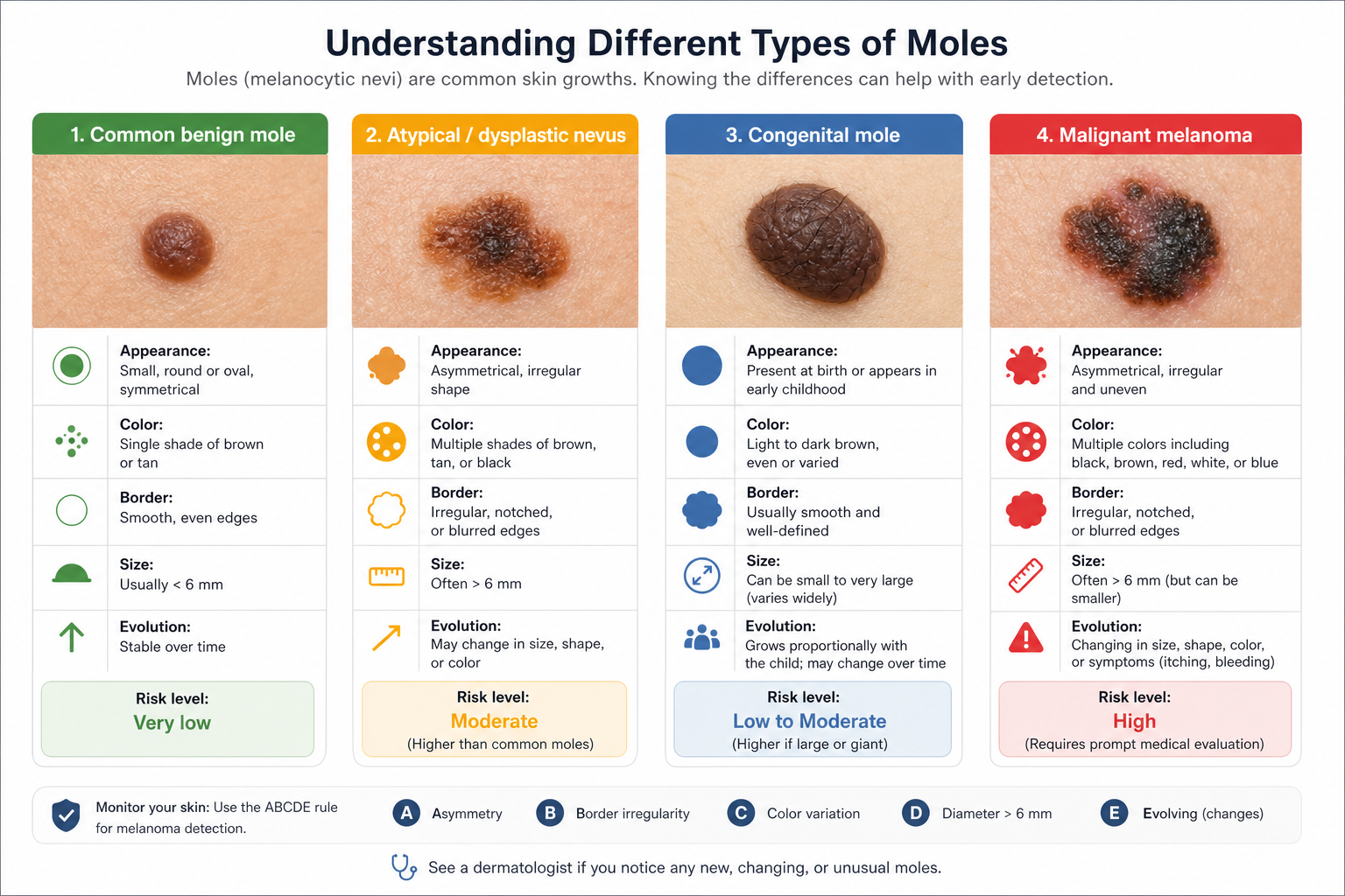
Not every mole requires removal, but certain features should prompt a professional assessment. The ABCDE rule is the most widely used clinical framework for identifying potentially suspicious moles:
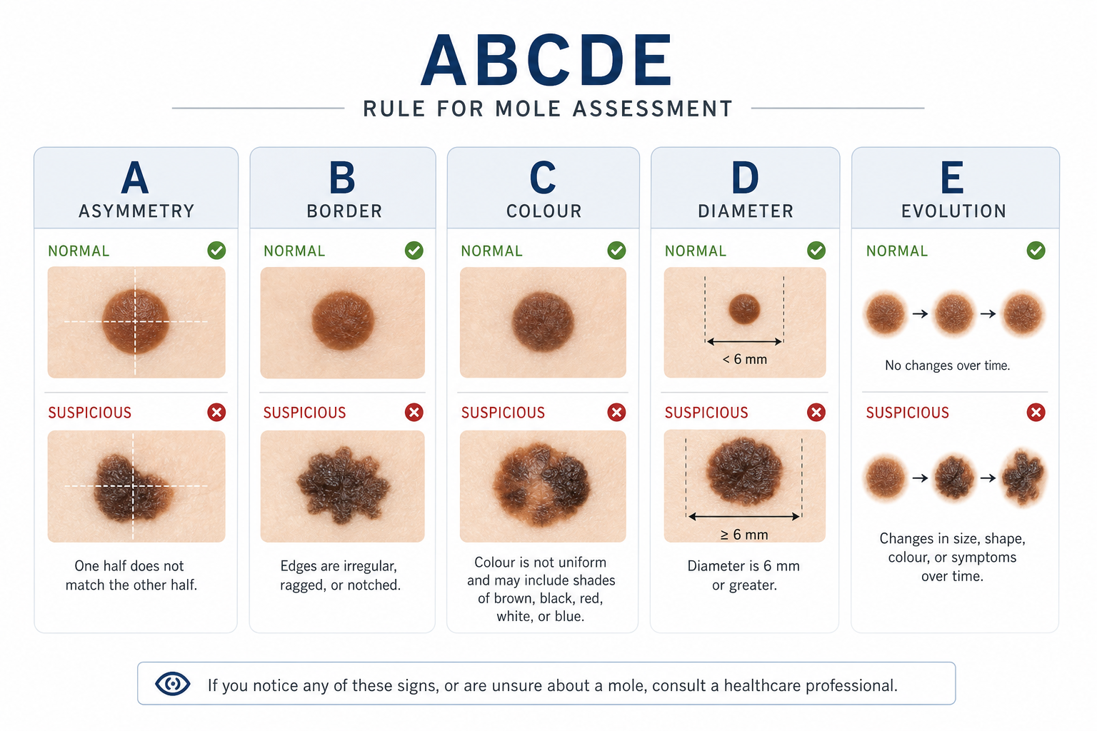
- Asymmetry: One half of the mole does not match the other in shape.
- Border: The edges are irregular, ragged, notched, or blurred rather than smooth and well-defined.
- Colour: The mole contains multiple shades — brown, black, red, white, or blue — rather than a uniform colour.
- Diameter: The mole is larger than approximately 6 mm (roughly the size of a pencil eraser), though melanomas can be smaller.
- Evolution: The mole has changed in size, shape, colour, or texture, or has developed new symptoms such as itching, bleeding, or crusting.
A complementary concept is the “ugly duckling” sign — a mole that simply looks different from all the others on your body. Most people’s moles share a general family resemblance; one that stands out as distinctly different warrants attention regardless of whether it meets every ABCDE criterion.
It is also worth noting that melanoma can present differently in people with darker skin tones. In these cases, melanoma may appear under the nails, on the palms of the hands, or on the soles of the feet, and may not display the classic ABCDE pattern as clearly. Anyone with concerns about a changing skin lesion, regardless of skin tone, should seek a professional assessment rather than relying solely on self-assessment tools.
Types of Moles and Their Associated Risk Levels
| Mole Type | Description | Cancer Risk | Typical Recommendation |
|---|---|---|---|
| Common benign mole (melanocytic nevus) | Uniform colour, smooth border, symmetrical, stable | Very low | Monitor; remove if changing or for cosmetic reasons |
| Atypical / dysplastic nevus | Irregular border, mixed colour, larger than average | Elevated; higher melanoma association | Professional assessment; often excised with margins |
| Congenital mole (present from birth) | Varies; large congenital nevi carry higher risk | Depends on size; large lesions carry higher risk | Dermatologist monitoring; removal considered for larger lesions |
| Malignant melanoma | Irregular, evolving, may bleed or crust | Malignant — requires treatment | Surgical excision with clear margins; oncology referral |
Professional Mole Removal: What the Procedure Involves
Professional mole removal is typically performed as an outpatient procedure under local anaesthetic, meaning the area is numbed and the patient remains awake and comfortable throughout. The method chosen depends on the size, depth, location, and clinical appearance of the mole, as well as whether the primary goal is diagnostic, cosmetic, or both.
Surgical Excision
Surgical excision involves cutting out the full thickness of the mole along with a margin of surrounding healthy tissue, then closing the wound with sutures. This method is preferred for suspicious or atypical moles because it provides the best chance of achieving clear surgical margins and yields a complete tissue specimen for pathological analysis. Recovery typically involves keeping the area clean and dry for a period determined by the clinician, with suture removal at a follow-up appointment.
Shave Excision
Shave excision removes the mole at or just below the skin surface using a surgical blade, without sutures. It is generally used for raised, benign-appearing moles and produces a flat wound that heals without stitches. While it is a quicker procedure with minimal downtime, shave excision carries a higher recurrence rate than surgical excision and may not be appropriate for moles with suspicious features, as it may not capture the deepest cells.
Punch Biopsy
A punch biopsy uses a circular cutting tool to remove a cylindrical core of skin. It is often used as a diagnostic tool to sample a portion of a larger or suspicious lesion before deciding on definitive treatment. The specimen is sent for pathological examination, and further treatment is planned based on the results.
Mohs Surgery
Mohs surgery is a specialized technique reserved for confirmed skin cancers, particularly in areas where tissue conservation is important, such as the face. The surgeon removes thin layers of tissue one at a time, examining each layer under a microscope before proceeding. This continues until no cancerous cells remain, achieving the highest possible cure rate while preserving as much healthy tissue as possible. Mohs surgery is not used for routine benign mole removal.
What Happens After Mole Removal
After any professional mole removal, the excised tissue should be sent to a pathology laboratory for histopathological analysis. A pathologist examines the cells under a microscope and produces a pathology report confirming whether the mole was benign, contained precancerous changes, or was malignant. This step is not optional for suspicious lesions — it is the mechanism by which any pre-existing cancer is identified and appropriate follow-up is arranged.
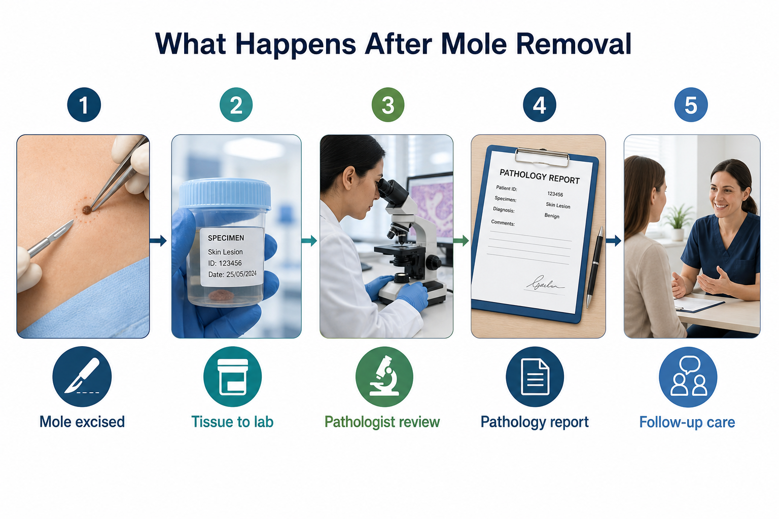
If the pathology report confirms a benign mole with clear margins, no further treatment is typically required, though regular skin checks are still recommended. If the report identifies an atypical dysplastic nevus with cells extending to the margin, re-excision with wider margins is usually advised. If malignant melanoma is confirmed, the patient will be referred for further management, which may include wider re-excision, sentinel lymph node assessment, or oncology review depending on the stage.
It is important to understand that removing one mole does not protect against new moles developing elsewhere, nor does it reduce the underlying skin cancer risk associated with UV exposure, genetic factors, or a high number of atypical moles. Ongoing sun protection, regular skin self-examination, and annual dermatologist visits remain relevant after mole removal.
Understanding Clear Surgical Margins
The concept of clear surgical margins is central to safe mole removal, yet it is rarely explained clearly to patients. When a surgeon removes a mole, they aim to excise not just the visible lesion but also a small border of surrounding normal-appearing tissue. This border is the surgical margin. “Clear margins” means that when the pathologist examines the edges of the removed specimen, no abnormal or cancerous cells are found at the boundary — indicating that the entire lesion has been removed.
“Positive margins” means abnormal cells are present at the edge of the specimen, suggesting that some cells may have been left behind in the patient’s skin. In this situation, further treatment — typically re-excision — is recommended to remove any remaining tissue. The width of margin recommended varies depending on the type of lesion: a benign mole may require only a narrow margin, while a confirmed melanoma requires wider margins determined by the tumour’s thickness. Understanding this concept helps patients appreciate why the choice of removal method and the skill of the practitioner directly affect the safety and completeness of the procedure.
Insurance and Cost Considerations
Whether mole removal is covered by health insurance or the NHS typically depends on the clinical reason for removal. Removal of a mole assessed as suspicious or potentially malignant is generally considered a medical procedure and is more likely to be covered or funded. Removal for purely cosmetic reasons — because a patient dislikes the appearance of a benign mole — is usually classified as a cosmetic procedure and is typically not covered by standard health insurance or public healthcare systems. If you are uncertain, it is worth checking with your insurer or healthcare provider before your appointment, as the clinical documentation from your dermatologist’s assessment may influence the decision.
Frequently Asked Questions
Does cutting into a mole make cancer spread?
No. There is no reliable clinical evidence that incising a mole during professional removal causes cancer to spread. The concern likely stems from confusion between correlation and causation — cancer diagnosed after removal was pre-existing, not caused by the procedure. For confirmed melanoma, surgical excision with adequate margins is the recommended treatment, not something to avoid.
If a mole grows back after removal, does that mean it is cancerous?
Not necessarily. Mole recurrence most commonly occurs after shave excision when some cells were left behind. The regrown tissue is often benign, though it should always be assessed by a dermatologist. In some cases, regrowth can display features that mimic melanoma under the microscope — a phenomenon called pseudomelanoma — which is why professional evaluation and pathological analysis of any recurrent lesion are important.
Is laser mole removal safe?
Laser removal can be used for certain superficial, benign-appearing moles for cosmetic purposes. However, laser treatment destroys the tissue rather than excising it, which means no specimen is available for pathological analysis. For this reason, laser removal is generally not recommended for moles with any suspicious features, and a clinical assessment should always precede any removal method to rule out malignancy first.
What should I do if I already removed a mole at home?
See a trained dermatologist as soon as possible. Even if the area appears healed, a skin specialist can examine the site for any remaining tissue, assess surrounding skin for suspicious changes, and advise on whether further evaluation is needed. The key concern is that no tissue was sent for pathological analysis, so any pre-existing abnormality in that mole cannot be retrospectively assessed.
How often should I have my skin checked after mole removal?
This depends on your individual risk profile, including your personal and family history of skin cancer, the number of atypical moles you have, and your history of UV exposure. As a general guide, an annual full-body skin check with a dermatologist is recommended for most adults, with more frequent checks for those at higher risk. Regular skin self-examination between appointments helps identify any new or changing lesions promptly.
Can a benign mole become cancerous over time?
Most common benign moles remain stable throughout a person’s life. However, any mole can theoretically undergo changes over time, particularly with cumulative UV exposure. Atypical or dysplastic nevi carry a statistically higher association with melanoma than common moles, though the absolute risk for any individual mole remains low. This is why monitoring for evolution — the “E” in the ABCDE rule — is important, and why any mole that changes should be assessed professionally rather than observed at home indefinitely.
When to Seek Professional Assessment About a Mole
The decision about whether and how to remove a mole should always begin with a professional assessment by a trained dermatologist or skin specialist. A clinician can examine the mole using dermoscopy — a magnified, illuminated view of the skin’s surface layers — to identify features not visible to the naked eye, and can advise on the most appropriate management based on the mole’s characteristics and your individual risk factors.
Seek assessment promptly if a mole is new and growing rapidly, if an existing mole has changed in size, shape, or colour, if a mole bleeds, itches, or crusts without an obvious cause, or if you simply have a mole that concerns you. Early assessment of a suspicious mole carries a far lower risk than delayed diagnosis. The evidence is clear: the danger lies not in professional mole removal, but in avoiding it when it is clinically indicated.