When to Get Moles Checked: A Practical Guide to Mole Monitoring
Most people have moles, and the majority are completely harmless. Knowing when to get moles checked by a qualified professional, however, is one of the most practical steps you can take for your long-term skin health. Changes in size, shape, colour, or texture can sometimes indicate that a mole warrants closer attention, and early assessment is consistently associated with better outcomes when any concern does turn out to be significant.
Key Takeaways
- Most moles are benign, but any mole that changes in appearance, bleeds, itches persistently, or looks noticeably different from your other moles should be assessed by a qualified clinician promptly.
- The ABCDE criteria — Asymmetry, Border, Colour, Diameter, and Evolving — provide a useful self-monitoring framework, but they are not a substitute for professional examination.
- Adults should consider a baseline full-body skin check, particularly if they have a large number of moles, a personal or family history of skin cancer, or significant past sun exposure.
- Dermoscopy and digital mole mapping are tools that allow clinicians to examine moles in greater detail than the naked eye alone permits.
- If you are ever uncertain about a mole, seeking professional assessment is always the appropriate course of action.
What Is a Mole and Why Do They Form?
A mole, known medically as a melanocytic naevus, is a growth on the skin formed by a cluster of pigment-producing cells called melanocytes. Melanocytes are responsible for producing melanin, the pigment that gives skin its colour. When these cells group together rather than spreading evenly throughout the skin, a mole forms. Most moles appear during childhood and adolescence, though new moles can develop in adulthood, and existing moles may change gradually over time in response to hormonal shifts, sun exposure, or ageing.
The average adult has between 10 and 40 moles, though some individuals have considerably more. Having a higher number of moles — generally considered to be more than 50 — is associated with a modestly increased risk of melanoma, the most serious form of skin cancer. This does not mean that having many moles is dangerous in itself, but it does make regular monitoring more important. Moles can be flat or raised, smooth or rough, and range in colour from pale pink to dark brown or black. Most remain stable throughout a person’s life.
The ABCDE Rule: A Framework for Self-Monitoring
The ABCDE criteria are a widely used self-examination tool that can help you identify moles that may need professional review. Understanding each element gives you a practical starting point for monitoring your own skin between clinical appointments.
- Asymmetry: A typical benign mole is roughly symmetrical — if you were to draw a line through the centre, both halves would look similar. Asymmetry, where one half looks noticeably different from the other, is worth having assessed.
- Border: Benign moles usually have smooth, well-defined edges. Irregular, ragged, notched, or blurred borders can be a reason to seek professional review.
- Colour: Most harmless moles are a single, uniform shade of brown. Variation in colour within a single mole — including patches of black, red, white, or blue — warrants attention.
- Diameter: Moles larger than approximately 6 millimetres across (roughly the size of a pencil eraser) are traditionally flagged for review, though smaller moles can also be significant if other features are present.
- Evolving: Any mole that is changing — in size, shape, colour, or texture — or that develops new symptoms such as itching, bleeding, or crusting should be assessed without delay.
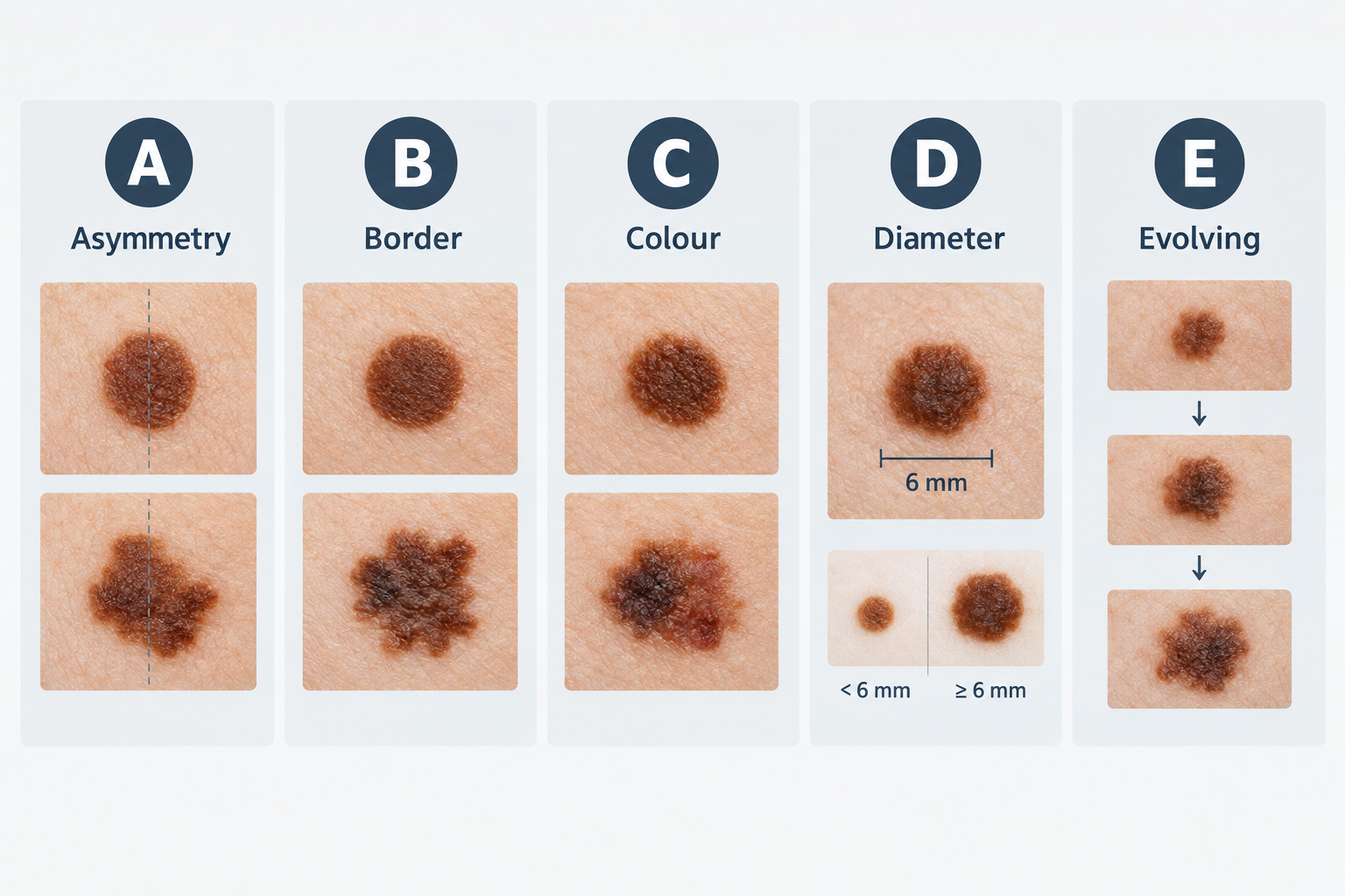
The ABCDE framework is a helpful guide, but it has limitations. Some melanomas do not display all of these features, and some benign moles may appear irregular without being dangerous. Professional assessment remains essential for any mole that concerns you, regardless of whether it fits neatly into these criteria.
Specific Warning Signs That Warrant Prompt Assessment
Beyond the ABCDE framework, certain specific changes or characteristics should prompt you to seek professional review without waiting for a routine appointment. These signs do not automatically indicate a serious problem, but they do mean that a qualified clinician should examine the mole in person.
- A mole that bleeds without being caught or scratched
- Persistent itching or tingling in or around a mole
- A mole that becomes raised, develops a lump within it, or changes texture noticeably
- A new dark streak appearing under a fingernail or toenail
- A sore or lesion that does not heal within a few weeks
- A mole that looks noticeably different from all your other moles — sometimes called the “ugly duckling” sign
- Any new growth on the skin that appears after the age of 40, particularly if it is dark, irregular, or growing
It is worth emphasising that many of these changes have benign explanations. A mole may bleed because it was caught on clothing, or it may itch due to dry skin. The point is not to cause alarm but to ensure that a professional can make that determination rather than leaving it to guesswork.
Who Should Consider Regular Mole Checks?
While anyone can develop a problematic mole, certain factors are associated with a higher risk of melanoma and other skin cancers. People in the following groups may benefit from more frequent or structured professional skin monitoring.
- Personal or family history of melanoma or skin cancer: A first-degree relative who has had melanoma increases your own risk, and a personal history of any skin cancer warrants ongoing surveillance.
- Large number of moles: Having more than 50 moles is associated with increased melanoma risk and makes thorough self-examination more challenging.
- Atypical or dysplastic moles: Some moles have irregular features under dermoscopic examination without being cancerous. These atypical naevi may require more frequent monitoring.
- Fair skin, light eyes, or red or blonde hair: These characteristics are associated with reduced natural protection against ultraviolet radiation and higher skin cancer risk.
- History of significant sun exposure or sunburn: Repeated sunburn, particularly during childhood, and prolonged cumulative sun exposure are established risk factors for melanoma.
- History of sunbed use: Regular use of ultraviolet tanning equipment is associated with increased melanoma risk, particularly when use began at a younger age.
- Immunosuppression: People taking immunosuppressant medications or living with conditions that affect immune function may have a higher risk of skin cancer and may benefit from more frequent skin checks.
- Large congenital moles: Moles present from birth, particularly larger ones, carry a different risk profile and are typically monitored from an early age.
If you are uncertain whether your personal history places you in a higher-risk group, a consultation with a dermatologist or a clinician experienced in skin cancer screening can help clarify what monitoring schedule may be appropriate for you.
How Often Should Moles Be Checked?
There is no single universally agreed interval for mole checks that applies to every person. General guidance from dermatology organisations suggests that adults at average risk should perform monthly self-examination of their skin and consider a professional full-body skin check at least once, ideally establishing a baseline record of their moles. For people with elevated risk factors, annual or more frequent professional checks are commonly recommended, though the appropriate interval depends on individual circumstances and should be discussed with a qualified clinician.
Self-examination is a valuable habit but has clear limitations. It is difficult to examine your own back, scalp, and other hard-to-see areas thoroughly. A partner or family member can help, but this is not a substitute for professional assessment using appropriate tools. Photographing your moles periodically — using consistent lighting and distance — can help you notice changes over time, though clinical-grade photography and digital mapping offer considerably greater precision.
What Happens During a Professional Mole Check?
A professional mole check typically begins with a discussion of your skin history, including any moles that concern you, your personal and family history of skin cancer, your sun exposure history, and any medications you take. The clinician will then examine your skin systematically, usually asking you to undress to allow a thorough full-body assessment. The examination covers areas that are easy to overlook during self-examination, including the scalp, between the toes, the soles of the feet, and the genital area.
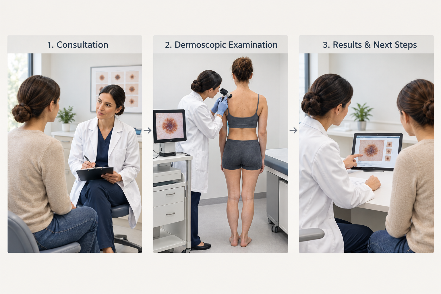
Most clinicians performing mole checks use a dermoscope, a handheld device that combines magnification with a light source to allow detailed examination of the skin’s surface and subsurface structures. Dermoscopy significantly improves the accuracy of mole assessment compared with the naked eye alone, allowing clinicians to identify features that are not visible without magnification. The examination itself is non-invasive and painless.
If a mole appears suspicious during examination, the clinician will discuss the findings with you and explain the recommended next steps. This may involve referral for a biopsy, in which a small sample of the mole is removed and sent for laboratory analysis. A biopsy is the only way to definitively determine whether a mole contains abnormal cells. Not every mole that is referred for biopsy turns out to be cancerous — many are removed as a precaution or because their features are ambiguous under dermoscopy.
Digital Mole Mapping: What It Involves
Digital mole mapping is a more comprehensive form of skin surveillance that uses standardised photography and, in some systems, dermoscopic imaging to create a detailed record of all moles on the body. This baseline record is then compared at subsequent appointments to identify any moles that have changed in appearance since the previous assessment.
Digital mole mapping is particularly useful for people with a large number of moles or atypical naevi, where tracking individual lesions over time would be difficult without a photographic reference. It allows clinicians to detect subtle changes that might not be apparent from a single examination. The technology does not replace clinical judgement — a trained clinician still interprets the images — but it adds an objective layer of comparison that can improve the detection of early changes.
Not all clinics offer digital mole mapping, and the availability and cost vary. If you have a high mole count or a history of atypical moles, it is worth asking whether this type of monitoring may be appropriate for your situation.
Moles in Children and Adolescents
Most moles in children are benign, and melanoma in children is rare. However, children can develop moles that warrant monitoring, and parents should be aware of the same general warning signs that apply to adults. Congenital melanocytic naevi — moles present at birth — vary considerably in size and appearance. Larger congenital naevi carry a different risk profile from moles that develop later in life and are typically assessed by a dermatologist to establish an appropriate monitoring plan.
New moles commonly appear during childhood and adolescence as a normal part of development. A mole that appears suddenly, grows rapidly, or displays irregular features in a child should be assessed by a clinician. Sun protection during childhood and adolescence is particularly important, as evidence consistently links early sunburn and cumulative ultraviolet exposure to increased lifetime melanoma risk.
Moles and Sun Protection
Ultraviolet radiation from sunlight and artificial sources such as sunbeds is the most significant modifiable risk factor for melanoma. Sun protection does not eliminate risk entirely, but consistent use of broad-spectrum sunscreen with an SPF of 30 or higher, protective clothing, and avoidance of peak ultraviolet hours can meaningfully reduce cumulative exposure. People with a large number of moles or other risk factors have particular reason to prioritise sun protection as part of their routine.
Existing moles can darken temporarily with sun exposure, which is a normal response to ultraviolet radiation. This temporary darkening is different from a persistent change in colour or the development of new colour variation within a mole. If you notice that a mole has changed colour and the change does not resolve after sun exposure has reduced, professional assessment is appropriate.
When a Mole Is Removed: What to Expect
If a clinician recommends removing a mole — either because it appears suspicious or because it is causing physical discomfort — the procedure is typically straightforward and performed under local anaesthetic. The most common method is surgical excision, in which the mole and a small margin of surrounding skin are removed and the wound is closed with sutures. The removed tissue is sent to a laboratory for histological analysis, which examines the cells under a microscope to determine whether any abnormality is present.
Recovery from mole removal is generally quick. The area may be tender for a few days, and a small scar is expected. The size and visibility of the scar depend on the location and size of the mole, the technique used, and individual healing characteristics. Results of the laboratory analysis are usually available within one to two weeks, after which the clinician will discuss the findings and any further steps if required.
Mole removal for purely cosmetic reasons — when a mole is not clinically suspicious but is unwanted — is a separate consideration. In this case, the decision involves weighing the appearance of the mole against the certainty of a scar, and the approach should be discussed carefully with a qualified clinician who can also confirm that the mole does not require removal for clinical reasons.
Frequently Asked Questions
Can a mole become cancerous overnight?
Melanoma develops over time rather than appearing overnight. However, some changes can seem to appear quickly, which is why any mole that appears to change rapidly should be assessed promptly rather than monitored at home for an extended period.
Is it normal for moles to itch occasionally?
Occasional mild itching around a mole can have benign causes such as dry skin or irritation from clothing. Persistent itching that does not resolve, or itching accompanied by other changes in the mole, warrants professional assessment.
Do all suspicious moles turn out to be cancerous?
No. Many moles that are referred for biopsy or removal turn out to be benign. Clinicians apply a cautious approach because early detection significantly improves outcomes when a mole does contain abnormal cells. A referral or recommendation for biopsy is not a diagnosis.
Can I use a smartphone app to check my moles?
Several smartphone applications use image analysis to assess moles, but their accuracy is variable and they are not a substitute for professional dermoscopic examination by a trained clinician. Apps may be useful as a prompt to seek professional review but should not be relied upon to rule out concern.
How long does a full-body mole check take?
A thorough full-body skin examination typically takes between 20 and 45 minutes, depending on the number of moles present and whether dermoscopy is used for individual lesions. Digital mole mapping appointments may take longer.
Should I be concerned about a mole that has been present since childhood?
A mole that has been stable since childhood and has not changed is generally reassuring. However, any mole — regardless of how long it has been present — should be assessed if it begins to change in appearance, bleeds, or develops other new features.
What is the difference between a mole and a seborrhoeic keratosis?
Seborrhoeic keratoses are common benign skin growths that can resemble moles but are not formed from melanocytes. They often have a waxy, stuck-on appearance and may be rough in texture. A clinician can usually distinguish between the two during examination, though dermoscopy helps confirm the diagnosis when there is uncertainty.
Making the Decision to Have Your Moles Assessed
The most practical takeaway from any discussion of mole monitoring is straightforward: if a mole concerns you, seek professional assessment. Uncertainty is not a reason to delay — it is precisely the reason to consult a qualified clinician who can examine the mole with appropriate tools and provide an informed opinion. Early assessment, when any concern does prove to be significant, consistently offers the best possible outcomes.
For people without specific concerns, establishing a baseline professional skin check and developing a habit of monthly self-examination are reasonable steps for most adults. Those with elevated risk factors — including a family history of melanoma, a large number of moles, significant past sun exposure, or a history of atypical naevi — should discuss an appropriate monitoring schedule with a dermatologist or experienced skin cancer screening clinician. Sun protection remains the most important modifiable factor in reducing long-term risk, and it is never too late to make it a consistent part of your routine.