Milia vs Xanthelasma: What Are Those White Spots Under Your Eyes?
White spots under the eyes are a common concern that patients bring to aesthetic and dermatology clinics, yet the two most frequent causes — milia and xanthelasma — are quite different in their origin, significance, and how they should be managed. Knowing which one you are dealing with matters, because the appropriate response, and in some cases the urgency of seeking medical advice, differs considerably between the two conditions.
Key Takeaways
- Milia are small, benign keratin-filled cysts that commonly appear under the eyes and on the cheeks; they are not medically significant but can be removed for cosmetic reasons.
- Xanthelasma are yellowish-white cholesterol deposits that form on or near the eyelids; they may indicate elevated blood lipid levels and warrant a medical assessment.
- The two conditions look similar at a glance but differ in colour, texture, location, and underlying cause — a trained clinician can usually distinguish them on examination.
- Neither condition resolves reliably on its own without treatment, and home removal attempts carry a real risk of scarring and infection.
- Treatment options exist for both, but the right approach depends on an accurate diagnosis, individual health factors, and realistic expectations about outcomes.
What Are Milia?
Milia are tiny, dome-shaped cysts that sit just beneath the surface of the skin. Each milium (the singular form) is a small pocket filled with keratin — the same fibrous protein that makes up hair and nails. They typically measure between one and three millimetres in diameter and appear as pearly white or cream-coloured bumps. The skin over a milium is smooth and intact, which is why they cannot be squeezed out like a conventional blackhead or whitehead.
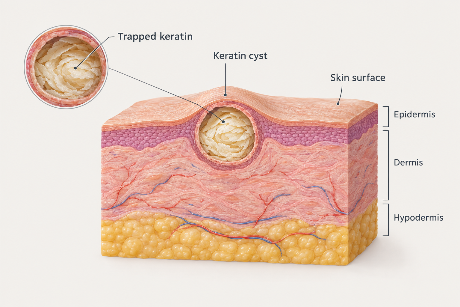
Milia form when dead skin cells become trapped in a small pocket near the skin’s surface rather than shedding normally. They can occur at any age, including in newborns, where they are extremely common and resolve without treatment within a few weeks. In adults, milia under the eyes and on the upper cheeks are particularly frequent because the skin in this area is thin, delicate, and prone to poor cell turnover. They can also develop after skin trauma, burns, blistering conditions, or prolonged use of heavy occlusive skincare products that prevent normal exfoliation.
Milia are entirely benign. They carry no medical risk, are not contagious, and are not associated with any systemic health condition. However, they tend to be persistent — adult milia rarely disappear without some form of intervention — and many patients find them cosmetically bothersome, particularly when they cluster around the under-eye area where the skin is already a focus of attention.
What Is Xanthelasma?
Xanthelasma palpebrarum is the medical term for soft, yellowish plaques that develop on or around the eyelids, most commonly near the inner corners. The word comes from the Greek for yellow skin, which reflects their characteristic colour. Unlike milia, xanthelasma are not cysts. They are deposits of lipid-laden macrophages — immune cells that have accumulated cholesterol and other fats — within the superficial layers of the skin.
Xanthelasma tend to be flat or only slightly raised, with a soft texture and a distinctly yellowish or cream-yellow hue that differs from the pure white of milia. They are usually larger than individual milia and may grow gradually over time. They most often appear on the upper eyelids but can also affect the lower eyelids and the skin immediately surrounding the eye area.
The clinical significance of xanthelasma extends beyond appearance. Approximately half of people with xanthelasma have elevated blood cholesterol or other lipid abnormalities, although xanthelasma can also occur in people with entirely normal lipid levels. Because of this association, a new diagnosis of xanthelasma should prompt a blood lipid profile test to rule out hyperlipidaemia or dyslipidaemia. Some research has also suggested a possible association between xanthelasma and an increased cardiovascular risk, though this relationship is not fully established and should be interpreted cautiously. A GP or physician is the appropriate first point of contact when xanthelasma is suspected.
How to Tell the Difference
Distinguishing milia from xanthelasma is usually straightforward for a trained clinician, though patients understandably find it difficult to tell them apart from photographs or self-examination. The key differences lie in colour, texture, location, and associated features.
| Feature | Milia | Xanthelasma |
|---|---|---|
| Colour | Pearly white or cream | Yellowish or cream-yellow |
| Texture | Firm, smooth dome | Soft, flat or slightly raised plaque |
| Size | 1–3 mm typically | Variable; often larger than milia |
| Location | Under eyes, cheeks, nose, forehead | Eyelids, especially inner corners |
| Cause | Trapped keratin | Cholesterol/lipid deposits |
| Medical significance | None | Possible lipid abnormality; warrants blood test |
| Tendency to grow | Stable or slowly increase in number | May enlarge gradually over time |
| Age of onset | Any age, including newborns | More common in middle age and older adults |
If you are uncertain which condition you have, a consultation with a dermatologist, GP, or experienced aesthetic practitioner is the appropriate first step. Self-diagnosis based on photographs alone is unreliable, and attempting to treat either condition at home without a confirmed diagnosis risks making the situation worse.
Causes and Contributing Factors
What Causes Milia to Form?
Primary milia develop spontaneously when keratin becomes trapped beneath the skin surface. Secondary milia can arise after skin injury or inflammation, including sunburn, dermabrasion, laser resurfacing, or blistering skin conditions such as epidermolysis bullosa or porphyria cutanea tarda. In the under-eye area specifically, the use of thick, occlusive eye creams or heavy moisturisers is a commonly cited contributing factor, as these products may impair the normal shedding of dead skin cells. Certain topical steroid preparations used around the eyes have also been associated with milia formation.
Genetics may play a role in susceptibility, and some individuals simply have skin that is more prone to poor keratin turnover in this area. Sun damage over time can also impair the skin’s natural exfoliation process, which may explain why milia become more common with age in some patients.
What Causes Xanthelasma to Develop?
Xanthelasma develop when lipid-laden cells accumulate in the dermis of the eyelid skin. The precise mechanism is not fully understood, but elevated circulating lipids — particularly low-density lipoprotein (LDL) cholesterol — are a recognised contributing factor in many cases. Conditions associated with secondary hyperlipidaemia, such as hypothyroidism, diabetes, liver disease, and certain kidney conditions, may also predispose individuals to xanthelasma. Some medications that affect lipid metabolism can be relevant as well.
However, xanthelasma can and does occur in people with completely normal lipid levels, which suggests that local tissue factors and individual susceptibility also play a part. The condition is more common in women and tends to appear in middle age or later, though it can occur earlier in people with familial hypercholesterolaemia — a genetic condition causing significantly elevated cholesterol from birth.
Treatment Options for Milia
Because milia are benign, treatment is entirely elective and driven by cosmetic preference. Several approaches are used in clinical practice, each with different suitability depending on the number of milia, their depth, and the patient’s skin type.
Manual Extraction
Manual extraction by a trained practitioner is one of the most straightforward approaches for individual or small clusters of milia. The practitioner makes a tiny incision in the skin overlying the milium using a sterile lancet or fine needle, then gently expresses the keratin contents. When performed correctly by a qualified professional, this technique is effective and leaves minimal marking. Attempting this at home is strongly discouraged — the skin around the eyes is exceptionally thin and delicate, and unskilled attempts frequently result in scarring, post-inflammatory hyperpigmentation, or infection.
Topical Retinoids
Topical retinoids, including tretinoin and over-the-counter retinol formulations, can help improve skin cell turnover and may gradually reduce the formation of new milia over time. They are generally more useful as a preventive measure or for patients with multiple milia than as a rapid treatment for existing lesions. Retinoids must be used with caution around the eye area due to the risk of irritation, and a practitioner’s guidance on appropriate formulation and application technique is advisable.
Chemical Exfoliation
Superficial chemical peels using alpha-hydroxy acids such as glycolic acid or lactic acid can support skin cell turnover and may help reduce milia over a course of treatments. These are typically used as part of a broader skin management plan rather than as a standalone milia treatment. Results vary, and the delicate periorbital skin requires careful product selection and application.
Laser and Energy-Based Treatments
Ablative laser treatments, including carbon dioxide laser and erbium laser, can be used to remove milia by precisely vaporising the cyst contents and overlying skin. Diathermy — the use of a fine electrical current — is another option used in some clinics. These approaches may be more suitable for patients with multiple or stubborn milia, but they carry a higher risk of post-treatment redness, temporary pigment changes, and, in less experienced hands, scarring. A thorough consultation and realistic expectations are essential before proceeding.
Treatment Options for Xanthelasma
Xanthelasma treatment involves two distinct considerations: addressing any underlying lipid abnormality through medical management, and removing the visible deposits for cosmetic or functional reasons. These are not mutually exclusive, and both may be relevant depending on the individual patient’s situation.
Medical Management of Underlying Lipid Levels
If a blood test reveals elevated cholesterol or other lipid abnormalities, addressing these through dietary changes, lifestyle modification, or lipid-lowering medication prescribed by a physician is an important first step. However, it is worth noting that treating elevated lipids does not reliably cause existing xanthelasma to resolve, and xanthelasma can recur after removal if lipid levels remain elevated. Medical management is primarily important for cardiovascular health rather than as a cosmetic treatment for the deposits themselves.
Trichloroacetic Acid (TCA) Application
Topical application of trichloroacetic acid at appropriate concentrations is a widely used clinic-based treatment for xanthelasma. The acid causes controlled destruction of the lipid deposits. Multiple sessions may be required, and there is a risk of pigmentation changes, particularly in patients with darker skin tones. Precise application is critical given the proximity to the eyes, and this procedure should only be performed by a practitioner experienced in periorbital treatments.
Surgical Excision
Surgical removal by a dermatologist or oculoplastic surgeon is an option for larger or more extensive xanthelasma. It offers the advantage of complete removal in a single procedure but carries the risk of scarring and, in some cases, ectropion — an outward turning of the eyelid — if too much skin is removed. Recurrence after surgical excision is possible, particularly if underlying lipid levels are not controlled.
Laser Treatment
Various laser modalities, including carbon dioxide laser, erbium laser, and pulsed dye laser, have been used to treat xanthelasma with varying degrees of success reported in the literature. Laser treatment can offer precise tissue removal with potentially less scarring than surgery in suitable cases, but recurrence remains a possibility. Evidence comparing different laser modalities for xanthelasma is limited, and outcomes depend significantly on lesion size, depth, and individual patient factors.
Cryotherapy
Cryotherapy — the application of liquid nitrogen to freeze and destroy tissue — has been used for xanthelasma, though it is generally considered less precise than laser or TCA approaches for this indication. It may be more suitable for smaller lesions and carries risks of pigmentation changes and blistering.
Risks and Limitations of Treatment
Both milia and xanthelasma treatments carry risks that patients should understand before proceeding. The periorbital area is anatomically sensitive, with thin skin, proximity to the eye itself, and a tendency toward post-inflammatory pigmentation changes, particularly in patients with medium to darker skin tones (Fitzpatrick skin types III–VI).
Recurrence is a meaningful limitation for both conditions. Milia can recur if the underlying cause — such as heavy skincare products or impaired skin turnover — is not addressed. Xanthelasma recurrence rates after treatment are reported to be significant, with some studies suggesting recurrence in a substantial proportion of patients within a few years, particularly when lipid levels remain elevated. Patients should be counselled about this possibility before committing to any removal procedure.
Scarring, though uncommon when treatment is performed by an experienced practitioner, is a real risk with any procedure involving the delicate eyelid skin. Post-inflammatory hyperpigmentation — darkening of the skin after treatment — is more common in patients with darker skin tones and may take months to resolve. Hypopigmentation, or lightening of the skin, is also possible with more aggressive treatments. Eye protection is essential during any laser or energy-based procedure in this area.
Who Should Seek a Medical Assessment First?
Anyone who notices new yellowish deposits around the eyelids and suspects xanthelasma should see a GP or physician before pursuing cosmetic removal. A fasting blood lipid profile is a straightforward test that can identify whether elevated cholesterol or triglycerides are present, and this information is important for overall health management regardless of any cosmetic treatment decision.

Patients with a personal or family history of cardiovascular disease, familial hypercholesterolaemia, diabetes, or thyroid conditions should be particularly prompt in seeking medical review if xanthelasma appears. In these cases, the skin finding may be a useful early indicator of a condition that benefits from medical attention.
Milia, by contrast, do not require urgent medical assessment. However, if you are unsure whether a lesion is a milium or something else — particularly if a spot is growing rapidly, changing colour, bleeding, or causing discomfort — a professional assessment is always the appropriate course of action. Not every white spot under the eye is a milium or xanthelasma, and a qualified practitioner can rule out other causes including syringomas (benign sweat duct tumours), sebaceous hyperplasia, or, in rare cases, more significant lesions.
Skincare and Prevention
Preventing milia from forming or recurring involves supporting normal skin cell turnover and avoiding factors that impair it. Incorporating a gentle exfoliating ingredient — such as a low-concentration retinol or a mild AHA — into a regular skincare routine may help, though care is needed around the eye area. Choosing non-comedogenic, lightweight eye creams rather than heavy occlusive formulations is a practical step for those prone to milia in this area. Consistent daily sun protection also supports healthy skin cell turnover and reduces cumulative photodamage that can impair the skin’s natural exfoliation process.
For xanthelasma, the most meaningful preventive measure — where relevant — is maintaining healthy lipid levels through diet, exercise, and medical management where prescribed. This does not guarantee that xanthelasma will not develop or recur, but it addresses the most modifiable contributing factor and has broader cardiovascular health benefits.
Frequently Asked Questions
Can I remove milia at home?
Home removal of milia is not recommended. The skin around the eyes is very thin and easily damaged. Attempting to pierce or squeeze milia without sterile equipment and proper technique frequently causes scarring, infection, or post-inflammatory pigmentation. Professional extraction by a trained practitioner is safer and more effective.
Do milia go away on their own in adults?
Unlike milia in newborns, which typically resolve within a few weeks, adult milia rarely disappear without some form of treatment or intervention. They may remain stable for months or years. Improving skin cell turnover through appropriate skincare can help prevent new ones from forming, but established milia generally require professional removal.
Is xanthelasma dangerous?
Xanthelasma itself is not dangerous in the direct sense — it does not damage the eye or surrounding tissue. However, its association with elevated blood lipids in a significant proportion of cases means it can be a useful indicator of an underlying condition that carries cardiovascular risk. Anyone with suspected xanthelasma should have their lipid levels checked by a doctor.
Will treating my cholesterol make xanthelasma disappear?
Lowering elevated cholesterol through medication or lifestyle changes does not reliably cause existing xanthelasma to resolve, though it may slow the development of new deposits and reduce the risk of recurrence after treatment. Medical management of lipid levels is important for health reasons but should not be expected to function as a cosmetic treatment for existing plaques.
Can xanthelasma come back after treatment?
Yes. Recurrence after treatment is a recognised limitation of all current xanthelasma removal methods, including laser, TCA application, and surgical excision. Recurrence is more likely when underlying lipid levels remain elevated. Patients should be informed of this possibility before deciding on treatment.
Are there other causes of white spots under the eyes I should know about?
Yes. Syringomas are benign tumours of the sweat ducts that can appear as small flesh-coloured or yellowish bumps around the lower eyelids and are sometimes mistaken for milia or xanthelasma. Sebaceous hyperplasia, closed comedones, and in rare cases more significant lesions can also present as spots in this area. A professional assessment is the only reliable way to confirm what you are dealing with.
How many sessions are needed to remove milia professionally?
Individual milia can often be extracted in a single appointment. Patients with multiple milia may require more than one session, and those using topical treatments such as retinoids to prevent recurrence should expect to use these consistently over weeks to months before seeing a meaningful reduction in new lesion formation.
Deciding What to Do About White Spots Under Your Eyes
The most important first step when you notice white spots under your eyes is to get an accurate diagnosis. Milia and xanthelasma may look superficially similar, but they have different causes, different implications for your health, and different treatment pathways. Treating one as if it were the other — or attempting home removal of either — is unlikely to produce a good outcome and may cause unnecessary harm to the delicate skin around the eyes.
If the spots are milia, you have the reassurance that they pose no health risk, and you can make an informed, unhurried decision about whether cosmetic removal is something you want to pursue. If the spots are xanthelasma, a visit to your GP for a lipid blood test is a sensible and straightforward next step, regardless of whether you ultimately choose to have the deposits removed.
For either condition, professional treatment in a clinical setting — by a dermatologist, GP with a special interest in skin, or an experienced aesthetic practitioner — offers meaningfully better outcomes and lower risk than any home remedy. A thorough consultation will allow a practitioner to confirm the diagnosis, discuss the most appropriate treatment options for your skin type and the extent of the lesions, and set realistic expectations about results, recovery, and the possibility of recurrence. If you are concerned about white spots under your eyes, booking a consultation with a qualified professional is a practical and proportionate response.