Types of Dark Circles: Pigmented vs Vascular — What’s the Difference?
Dark circles under the eyes are one of the most common cosmetic concerns brought to aesthetic clinics, yet they are frequently misunderstood — and, as a result, frequently mistreated. The term “dark circles” covers several distinct conditions with different underlying causes, and the most important distinction is between pigmented dark circles and vascular dark circles. Identifying which type you have is the essential first step, because a treatment that works well for one type may produce little or no improvement for another.
Key Takeaways
- Dark circles are not a single condition. Pigmented and vascular dark circles have different causes, appearances, and treatment pathways.
- Pigmented dark circles involve excess melanin in the skin, while vascular dark circles are caused by visible blood vessels or blood pooling beneath thin under-eye skin.
- A third category — structural dark circles — results from volume loss, hollowness, or shadowing, and often overlaps with the other two types.
- Accurate identification of the type, or combination of types, is essential before any treatment is considered.
- No single treatment corrects every type of dark circle, and results vary considerably between individuals.
Why Identifying the Type of Dark Circle Matters
The under-eye area is anatomically complex. The skin here is among the thinnest on the body — typically around 0.5 mm compared with 2 mm elsewhere on the face — which means that changes in pigmentation, blood flow, or underlying tissue structure become visible more readily than in other areas. When a patient presents with dark circles, a clinician needs to determine whether the discolouration originates in the skin itself, in the blood vessels beneath it, in the deeper soft tissue, or in some combination of all three. Treating a vascular problem with a pigment-targeting laser, for example, is unlikely to produce meaningful improvement and may cause unnecessary side effects.
Understanding the cause also helps set realistic expectations. Some types of dark circles respond well to topical skincare or in-clinic treatments; others are strongly influenced by genetics, anatomy, or lifestyle factors that cannot be fully corrected by any aesthetic intervention. A thorough clinical assessment — ideally including a simple stretch test and examination under different lighting conditions — is the starting point for any responsible treatment plan.
Pigmented Dark Circles: Causes and Characteristics
Pigmented dark circles occur when there is an excess of melanin — the pigment that gives skin its colour — in the skin of the lower eyelid and periorbital area. This type tends to produce a brown, tan, or greyish-brown discolouration that sits within the skin itself rather than beneath it. Pigmented dark circles are particularly common in people with Fitzpatrick skin types III to VI, meaning medium to darker skin tones, though they can affect any skin type.
What Causes Pigmented Dark Circles?
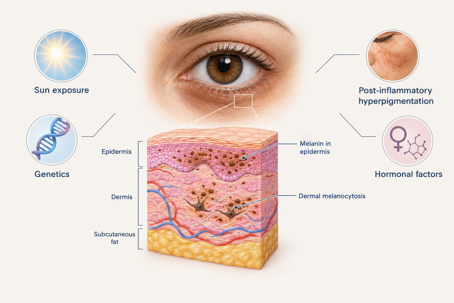
Several mechanisms can drive excess melanin production in the periorbital region. Post-inflammatory hyperpigmentation is one of the most frequent contributors: repeated rubbing of the eyes, allergic reactions, eczema, or contact dermatitis can trigger melanocyte activity and leave lasting pigmentation. Sun exposure is another significant factor, as ultraviolet radiation stimulates melanin production, and the under-eye area is often inadequately protected by sunscreen. Hormonal influences — including pregnancy, oral contraceptive use, and thyroid dysfunction — can also increase periorbital pigmentation in susceptible individuals. In some cases, the pigmentation is constitutional, meaning it is largely determined by genetics and has been present since childhood or early adulthood.
A less commonly discussed cause is dermal melanocytosis, where melanin-containing cells are located deeper within the dermis rather than in the superficial epidermis. This produces a blue-grey rather than brown discolouration and is more resistant to topical treatments because the pigment sits below the reach of most skincare ingredients.
How to Identify Pigmented Dark Circles
The stretch test is a useful clinical tool. When the skin beneath the eye is gently stretched, pigmented dark circles remain visible because the discolouration is within the skin itself. The colour does not significantly lighten or disappear with stretching. Under natural daylight, pigmented circles typically appear brown or tan. Wood’s lamp examination — a UV light used in dermatology — can help distinguish epidermal pigmentation (which fluoresces more clearly) from dermal pigmentation, which is deeper and harder to treat.
Vascular Dark Circles: Causes and Characteristics
Vascular dark circles arise from the visibility of blood vessels or haemoglobin-related pigments beneath the thin skin of the lower eyelid. Rather than a brown or tan tone, vascular dark circles typically appear blue, purple, pink, or reddish, depending on the depth and oxygenation of the underlying vessels. This type is extremely common and is often the dominant cause of dark circles in people with fair or thin skin.
What Causes Vascular Dark Circles?
The primary driver is the translucency of the periorbital skin. When the dermis is thin and the subcutaneous fat layer beneath the eye is minimal, the dense network of capillaries and venules in this region becomes visible through the skin surface. Fatigue and sleep deprivation are well-known aggravating factors: poor sleep causes vasodilation and increased blood pooling in the periorbital vessels, intensifying the bluish or purplish appearance. Allergies and chronic nasal congestion can impair venous drainage from the periorbital area, contributing to the same effect — a phenomenon sometimes called the “allergic shiner.”
Ageing plays a significant role in vascular dark circles. As the skin loses collagen and elastin over time, it becomes progressively thinner and more translucent, making underlying vessels more visible. Simultaneously, the fat pads that cushion the lower eyelid area diminish and descend, reducing the natural barrier between the skin surface and the vascular structures beneath. This is why vascular dark circles often worsen with age even in people who did not have them in youth.
Haemosiderin deposition is another vascular mechanism worth understanding. When small capillaries leak red blood cells into the surrounding tissue, the breakdown of haemoglobin produces haemosiderin — an iron-containing pigment that creates a brownish-red or rust-coloured discolouration. This can give vascular dark circles a mixed appearance that overlaps with pigmented types, making accurate diagnosis more challenging.
How to Identify Vascular Dark Circles
The stretch test is again informative. When the under-eye skin is gently stretched, vascular dark circles tend to lighten noticeably or temporarily disappear, because stretching the skin reduces the apparent density of the underlying vessels. The colour is typically blue, purple, or pinkish rather than brown. Vascular dark circles often appear worse after a poor night’s sleep, after alcohol consumption, or during allergy season — all situations that increase periorbital blood flow or impair drainage.
Structural Dark Circles: The Third Type
While pigmented and vascular dark circles are the two primary categories, a third type — structural or shadow-based dark circles — deserves attention because it is frequently overlooked and is often the dominant concern in older patients. Structural dark circles are not caused by pigmentation or blood vessel visibility but by the three-dimensional shape of the under-eye area. Volume loss in the tear trough region creates a hollow or concave contour that casts a shadow, producing the appearance of darkness even when the skin itself has normal colour and the underlying vessels are not unusually visible.
The tear trough is the groove that runs from the inner corner of the eye downward and outward along the junction between the lower eyelid and the cheek. As facial fat compartments diminish with age and the overlying skin loses support, this groove deepens and the shadow it creates becomes more pronounced. Structural dark circles are typically most visible in overhead or directional lighting and may appear less prominent in diffuse or frontal light. They do not respond to skincare, pigment-targeting treatments, or vascular lasers, but may improve with volume restoration using dermal fillers or, in some cases, surgical approaches.
Mixed-Type Dark Circles: The Most Common Presentation
In clinical practice, pure single-type dark circles are less common than mixed presentations. Many patients have elements of pigmentation, vascular visibility, and structural shadowing occurring simultaneously, and the relative contribution of each component varies between individuals. A person with naturally thin, fair skin may have prominent vascular dark circles that are worsened by haemosiderin deposition from minor capillary leakage, combined with early tear trough hollowing that adds a shadow component. A patient with a darker skin tone may have constitutional periorbital pigmentation alongside age-related volume loss.
This complexity is one reason why a single-product or single-treatment approach rarely produces satisfactory results for all patients. A clinician experienced in periorbital assessment will typically evaluate all three components and discuss a treatment plan that addresses the most significant contributing factors in the correct sequence.
Treatment Options by Dark Circle Type
Treatment selection depends directly on the identified type or combination of types. The following overview covers the main approaches used in aesthetic medicine, along with their realistic scope and limitations.
Treatments for Pigmented Dark Circles
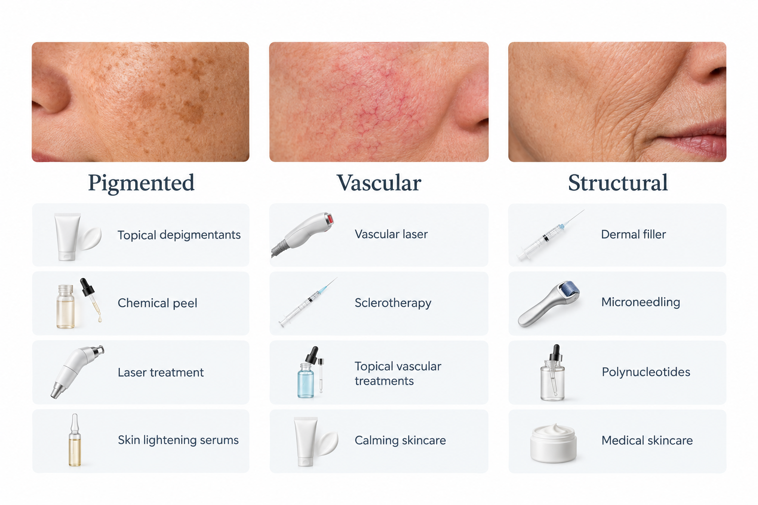
Topical depigmenting agents are often the first-line approach for epidermal pigmentation. Ingredients with established evidence for reducing melanin production include hydroquinone (where available and appropriately prescribed), azelaic acid, kojic acid, tranexamic acid, niacinamide, and vitamin C derivatives. These agents work by inhibiting tyrosinase — the enzyme responsible for melanin synthesis — or by interfering with melanin transfer to skin cells. Results are gradual, typically requiring consistent use over several months, and maintenance is usually necessary to prevent recurrence, particularly with ongoing sun exposure.
Topical retinoids, including tretinoin, can improve epidermal pigmentation by accelerating cell turnover and dispersing melanin granules. However, retinoids must be used cautiously in the periorbital area due to the risk of irritation, and they are not appropriate for everyone. Sun protection is an essential adjunct to any pigmentation treatment: without consistent broad-spectrum SPF use, pigmentation is likely to recur or worsen regardless of other interventions.
For more resistant epidermal pigmentation, in-clinic options include chemical peels using agents such as glycolic acid, lactic acid, or mandelic acid, which accelerate surface cell turnover. Laser treatments — particularly Q-switched Nd:YAG lasers and picosecond lasers — can target melanin in the skin, though these require careful patient selection, especially in darker skin tones where there is a risk of post-inflammatory hyperpigmentation. Intense pulsed light (IPL) may also be considered for superficial pigmentation in suitable candidates. Dermal pigmentation (melanocytosis) is significantly harder to treat and may not respond adequately to surface-level interventions.
Treatments for Vascular Dark Circles
Vascular dark circles are among the more challenging types to treat because the underlying cause — thin, translucent skin overlying a dense vascular network — is partly anatomical and partly age-related. Topical skincare has a limited role here, though ingredients that support collagen production (such as retinoids and vitamin C) may gradually improve skin thickness and reduce translucency over time. Caffeine-containing eye creams are widely marketed for their purported ability to constrict periorbital blood vessels and reduce puffiness, though clinical evidence for a meaningful or lasting effect on vascular dark circles is limited.
Vascular lasers and light-based devices — including pulsed dye lasers and certain IPL settings — can target haemoglobin in superficial blood vessels, reducing their visibility. These treatments may be helpful for patients whose vascular dark circles are driven primarily by superficial vessel prominence or haemosiderin deposition. Multiple sessions are typically required, and results vary depending on vessel depth and skin type.
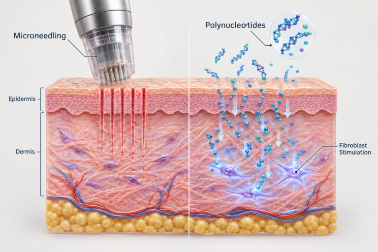
Polynucleotide injections (PDRN or PN treatments) have attracted growing interest as a treatment for the periorbital area. Polynucleotides are fragments of purified DNA that are thought to stimulate fibroblast activity, support collagen synthesis, and improve skin hydration and quality. By gradually thickening and improving the quality of the under-eye skin, polynucleotide treatments may reduce the translucency that makes vascular dark circles visible. Evidence in this specific application is still developing, and results are typically gradual, appearing over several weeks to months after a course of treatments. Polynucleotide injections do not directly target blood vessels or pigmentation, so their benefit is most relevant where thin skin quality is a primary contributing factor.
Skin boosters — injectable hyaluronic acid preparations designed to improve hydration and skin quality rather than add volume — may similarly improve the appearance of vascular dark circles by enhancing skin thickness and luminosity over time. Again, these are not direct vascular treatments, and realistic expectations should be discussed with a qualified practitioner.
Treatments for Structural Dark Circles
Structural dark circles caused by tear trough hollowing and volume loss are most effectively addressed by restoring the lost volume. Hyaluronic acid dermal fillers placed in the tear trough region can soften the shadow-creating groove and improve the transition between the lower eyelid and the cheek. Tear trough filler is a technically demanding procedure due to the proximity of important anatomical structures, the thinness of the overlying skin, and the risk of complications including bruising, swelling, the Tyndall effect (a bluish discolouration caused by superficially placed filler), and, in rare cases, vascular occlusion. It should only be performed by a clinician with specific training and experience in periorbital anatomy.
In patients with significant fat pad descent or excess lower eyelid skin, surgical options such as lower blepharoplasty may be more appropriate than injectable treatments. A surgical consultation is warranted when the structural component is pronounced and non-surgical approaches are unlikely to achieve a satisfactory result.
Lifestyle Factors That Affect All Types of Dark Circles
Regardless of the underlying type, several lifestyle factors can worsen dark circles and should be addressed alongside any clinical treatment. Chronic sleep deprivation increases periorbital vascular congestion and reduces skin repair processes, worsening both vascular and structural dark circles. Alcohol consumption causes vasodilation and fluid retention, temporarily intensifying the appearance of dark circles. Smoking accelerates collagen breakdown and impairs microcirculation, contributing to skin thinning and vascular visibility over time.
Allergic rhinitis and chronic nasal congestion impair venous drainage from the periorbital area and are a frequently underappreciated cause of persistent vascular dark circles. Managing underlying allergies — through antihistamines, nasal corticosteroids, or allergen avoidance — can produce a meaningful improvement in dark circle appearance that no cosmetic treatment alone would achieve. Adequate hydration supports skin quality generally, and a diet rich in antioxidants may help protect periorbital skin from oxidative stress, though the direct effect on established dark circles is modest.
Who Should Assess Your Dark Circles?
Because dark circles have multiple possible causes that require different treatment approaches, assessment by a qualified medical or aesthetic practitioner is strongly recommended before pursuing any in-clinic treatment. A dermatologist, plastic surgeon, or experienced aesthetic physician can perform a structured evaluation that distinguishes between pigmented, vascular, and structural components, identifies any underlying medical contributors such as thyroid disease or allergic conditions, and recommends a treatment plan appropriate to the individual’s skin type, anatomy, and goals.
Self-diagnosis based on appearance alone is unreliable, and purchasing treatments — whether topical or injectable — without a proper assessment risks wasting money on ineffective approaches or, in the case of injectable treatments, potential harm from procedures performed without adequate clinical justification or expertise.
Frequently Asked Questions
Can I tell at home whether my dark circles are pigmented or vascular?
The stretch test offers a useful clue: gently pull the skin beneath your eye taut. If the darkness remains largely unchanged, pigmentation is more likely. If it lightens noticeably, a vascular component is more probable. However, many people have mixed-type dark circles, and a clinical assessment is more reliable than self-testing alone.
Do dark circles get worse with age?
For most people, yes. Skin becomes thinner and loses collagen over time, making vascular structures more visible. Fat pads beneath the eye diminish and descend, deepening the tear trough shadow. Cumulative sun exposure can worsen pigmentation. The relative contribution of each factor varies between individuals.
Can eye creams genuinely improve dark circles?
Some topical ingredients — including vitamin C, niacinamide, retinoids, and tranexamic acid — have evidence supporting modest improvement in periorbital pigmentation with consistent long-term use. The effect on vascular or structural dark circles is generally limited. No over-the-counter eye cream can replicate the results of in-clinic treatments for significant dark circles.
Is tear trough filler safe for dark circles?
Tear trough filler can be effective for structural dark circles caused by volume loss, but it carries specific risks in this anatomically sensitive area, including bruising, swelling, the Tyndall effect, and rare but serious vascular complications. It is not appropriate for all types of dark circles and should only be performed by a clinician with relevant training and experience in periorbital anatomy.
How many sessions of polynucleotide treatment are typically needed for under-eye concerns?
Most protocols involve a course of two to four sessions spaced several weeks apart, followed by maintenance treatments. Results develop gradually over weeks to months as skin quality improves. Individual response varies, and polynucleotides are most relevant where skin thinning and quality are primary contributing factors rather than deep pigmentation or significant structural hollowing.
Can dark circles be permanently removed?
Permanent elimination is rarely achievable. Constitutional pigmentation and anatomical factors such as skin translucency and tear trough depth are largely determined by genetics and ageing processes that continue over time. Treatments can produce meaningful and sometimes long-lasting improvement, but maintenance is usually required, and results vary considerably between individuals.
Choosing the Right Approach for Your Type of Dark Circle
The most important step in addressing dark circles is not choosing a treatment — it is understanding which type of dark circle you have. Pigmented dark circles, driven by excess melanin in the skin, respond to a different set of interventions than vascular dark circles, which arise from the visibility of blood vessels through thin periorbital skin. Structural dark circles, caused by volume loss and shadowing in the tear trough, require a different approach again. Many people have a combination of all three, which is why a one-size-fits-all treatment rarely delivers satisfying results.
A structured clinical assessment by a qualified practitioner remains the most reliable way to identify the contributing factors in your specific case and to develop a treatment plan that is proportionate, evidence-informed, and appropriate for your skin type and anatomy. Whether the recommended approach involves topical skincare, light-based treatments, injectable polynucleotides, dermal fillers, or a combination of these, the outcome is most likely to be satisfying when it is based on an accurate diagnosis rather than a generic assumption about what dark circles are and what causes them. If you are considering treatment, a consultation with an experienced aesthetic or dermatology practitioner is a sensible and worthwhile first step.