What Is Vascular Occlusion After Filler? Signs, Risks & Treatment
Vascular occlusion after filler is a rare but serious complication of dermal filler injection procedures. It occurs when filler blocks blood flow within a blood vessel, reducing the blood supply to surrounding tissues. Without prompt treatment, tissue damage may develop and, in severe cases, skin necrosis or blindness can occur.
Although vascular occlusion is uncommon in aesthetic medicine, early identification is essential. Key warning signs include sudden pain, skin blanching, delayed capillary refill, unusual bruising, colour changes, and cool skin. Rapid assessment and management may significantly improve outcomes following filler treatment.




Thousands of satisfied customers

Vascular Occlusion Definition
Vascular occlusion refers to a blockage that disrupts normal blood flow through a vessel following a cosmetic injection. It is considered one of the most significant dermal filler complications because tissue survival depends on a continuous blood supply.
Unlike expected post-treatment reactions such as mild swelling or a bruise, vascular occlusion involves compromised circulation. Understanding the difference between normal recovery and a warning sign of compromise is important for both patients and practitioners.
Meaning Of Vascular Occlusion In Cosmetic Procedures
In aesthetics, vascular occlusion occurs when a blood vessel becomes obstructed, preventing oxygen-rich blood from reaching tissues. The resulting lack of blood supply may lead to tissue ischemia and, if untreated, tissue death.
Healthy circulation is essential because skin, fat, and other facial structures rely on constant oxygen delivery. Even short periods of impaired blood flow can affect tissue health in highly vascular areas of the face.
How Dermal Fillers Can Block Blood Flow
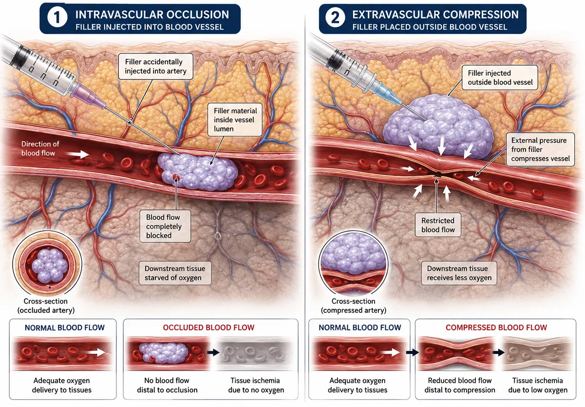
A dermal filler may enter an artery during injection or place pressure on a nearby blood vessel. Both mechanisms can reduce circulation and create a blockage within the facial vascular network.
The risk varies according to anatomy, treatment area, injection technique, filler material, and the type of filler used. Certain facial regions contain critical arterial pathways that require particular caution.
Difference Between Vascular Occlusion And Normal Filler Reactions
Normal filler injections commonly cause temporary swelling, tenderness, redness, and bruising. These effects typically improve gradually over several days and do not significantly affect blood flow.
Signs of vascular occlusion are different. Sudden severe pain, blanch, delayed capillary refill, unusual colour changes, or worsening symptoms following treatment require immediate clinical assessment rather than routine observation.
Normal Filler Reaction | Possible Vascular Occlusion |
Mild bruise | Rapid blanching |
Temporary swelling | Delayed capillary refill |
Mild tenderness | Significant pain |
Improves with time | Symptoms worsen |
Normal skin temperature | Cool skin |
How Vascular Occlusion Happens After Filler
Vascular occlusion following filler treatment may occur through several mechanisms. Understanding how complications develop helps explain why prevention and management protocols are emphasised throughout aesthetic medicine.
Intravascular Filler Injection
An intravascular injection occurs when filler is accidentally injected directly into a blood vessel. This can obstruct blood flow immediately and may create vascular compromise within the affected territory.
Even experienced injectors remain aware of this possibility because facial anatomy varies between individuals. Thorough anatomical knowledge remains a core safety requirement.
External Compression Of Blood Vessels
Not all cases of vascular occlusion result from direct vessel entry. Filler volume may compress nearby arteries or veins from outside the vessel wall.
External compression can restrict circulation, particularly in confined anatomical spaces where tissue expansion is limited.
Blood Flow Interruption And Tissue Ischemia
When blood flow is interrupted, tissues receive less oxygen and fewer nutrients. This process is known as tissue ischemia.
If ischemia persists, tissue damage may progress to skin necrosis. Early diagnosis and prompt treatment are therefore essential components of managing dermal filler complications.
Retrograde And Anterograde Filler Movement
In rare circumstances, filler may travel within vascular pathways. Retrograde movement occurs against normal blood flow, while anterograde movement follows circulation.
These mechanisms help explain severe complications reported in aesthetic medicine, including rare cases of vascular occlusion affecting vision.
Areas Most Vulnerable To Vascular Occlusion
Certain treatment areas are associated with a higher risk of vascular occlusion because of complex facial vascular anatomy and connections between vessels.
Nose
Non-surgical rhinoplasty involves filler injection near important arterial structures. Because the nose has limited collateral circulation, vascular compromise may lead to significant tissue damage if left untreated.
Glabella
The glabella, located between the eyebrows, contains vessels linked to the ophthalmic circulation. This region is frequently discussed in relation to severe complications.
Nasolabial Folds
Nasolabial fold treatments are common, but nearby arterial branches require careful injection technique and awareness of anatomical variation.
Lips
The lip area is highly vascular. Lip filler procedures, therefore, require careful planning, conservative placement, and continuous monitoring for signs of vascular occlusion.
Tear Trough Area
The tear trough region contains delicate structures and vascular connections near the eye. Specialist assessment is often important before treatment.
Forehead And Temple Regions
The forehead and temples contain major vessels that communicate with deeper vascular networks. Although complications remain uncommon, outcomes can be serious.
Treatment Area | Relative Risk Considerations |
Nose | Limited collateral blood supply |
Glabella | Connection to eye circulation |
Lips | Highly vascular tissue |
Tear trough | Sensitive anatomy |
Forehead | Important arterial pathways |
Temples | Complex vascular network |

Early Signs Of Vascular Occlusion
Recognising symptoms quickly may reduce the risk of tissue necrosis and long-term complications.
Sudden Pain During Injection
A sudden increase in pain during injection can be a warning sign. Although discomfort is expected with cosmetic procedures, severe pain may indicate compromised circulation.
Skin Blanching
Blanching describes whitening of the skin caused by reduced blood flow. It is often one of the earliest signs and symptoms of vascular compromise.
Delayed Capillary Refill
Capillary refill time assesses how quickly blood returns after gentle pressure on the skin. Delayed capillary refill suggests impaired circulation.
Livedo Reticularis Pattern
A mottled, net-like discolouration known as livedo reticularis may indicate disruption of blood flow through affected vessels.
Coolness Of Skin
Skin temperature can change when circulation decreases. Cool skin in the treatment area may be another warning sign requiring immediate review.
Common early warning signs:
- Sudden pain
- Skin blanch
- Delayed capillary refill
- Cool skin
- Livedo reticularis
- Rapid colour changes
Symptoms Of Progressive Vascular Occlusion
Progressive vascular occlusion may lead to worsening tissue ischemia if blood flow is not restored promptly.
Increasing Discoloration
Skin may transition from pale to purple, blue, or dark discolouration as oxygen deprivation worsens.
Persistent Pain
Pain often becomes more severe and prolonged as tissue damage develops.
Tissue Breakdown
Early tissue damage may appear as blistering, ulceration, or breakdown of the skin surface.
Skin Necrosis
Skin necrosis refers to tissue death caused by prolonged lack of blood supply. This is among the most serious filler complications.

Diagnosis Of Vascular Occlusion
Diagnosis relies primarily on clinical assessment and recognition of characteristic symptoms.
Clinical Assessment
Practitioners evaluate pain, colour changes, capillary refill, temperature differences, and the distribution of affected tissue.
Evaluation Of Blood Flow
Assessment commonly includes capillary refill testing and examination of blood flow patterns across the treatment area.
Differentiating Vascular Occlusion From Bruising
A bruise generally develops gradually and does not impair circulation. Vascular occlusion often presents with blanching, delayed capillary refill, and pain.
Importance Of Early Recognition
Early identification and treatment are consistently associated with improved outcomes and reduced risk of tissue necrosis.
Treatment For Vascular Occlusion After Filler
Management of vascular complications aims to restore circulation as quickly as possible.
Immediate Cessation Of Injection
The first step is to stop the injection immediately when vascular occlusion is suspected.
Hyaluronidase Administration
Hyaluronidase can dissolve the filler when a hyaluronic acid filler has been used. This enzyme plays a central role in managing vascular occlusion caused by hyaluronic acid products.
Massage And Warm Compresses
Massage and warm compress measures may be used to encourage blood flow and support circulation alongside other treatments.
Aspirin And Blood Flow Support
Some protocols discuss aspirin and additional supportive measures. Their use depends on practitioner judgement, patient factors, and current guidance.
Follow-Up Monitoring
Careful follow-up monitoring helps assess recovery, identify ongoing ischemia, and determine whether further intervention is required.
Management may include:
- Stop filler injections
- Hyaluronidase administration
- Massage
- Warm compress application
- Ongoing vascular assessment
- Follow-up reviews

Recovery Process And Healing Timeline
Recovery varies according to the severity of vascular compromise and the speed of intervention.
First 24 Hours
The initial treatment period focuses on restoring blood flow, monitoring symptoms, and identifying any progression.
Recovery During First Week
Many cases of vascular occlusion show gradual improvement during the first week when managed promptly.
Long-Term Healing
More severe tissue damage may require longer healing periods and can result in scarring or skin texture changes.
Factors Affecting Recovery
Important factors include:
- Timing of treatment
- Severity of blockage
- Treatment area involved
- Type of filler
- Individual healing response
- Quality of management
Potential Long-Term Effects
Although many cases recover well, some individuals experience lasting effects:
- Scarring may occur when tissue necrosis develops before circulation is restored.
- Changes in texture can result from tissue injury and healing processes.
- Hyperpigmentation and hypopigmentation may occur after significant inflammation or tissue damage.
- Complications can affect confidence and wellbeing, particularly when visible facial changes occur.
Prevention Of Vascular Occlusion
Prevention and management strategies are central to safe aesthetic medicine practice.
Detailed Knowledge Of Facial Anatomy
Understanding facial anatomy helps practitioners identify high-risk areas and avoid critical vessels.
Safe Injection Techniques
Common approaches include:
- Slow injection technique
- Low-pressure injection
- Small treatment volumes
- Continuous assessment
- Conservative placement
Appropriate Filler Selection
The filler used should be appropriate for the treatment area and patient characteristics.
Cannula Versus Needle Use
A cannula may reduce vessel penetration risk in some situations, although neither cannula nor needle use completely eliminates risk.
Patient Assessment Before Treatment
Assessment often includes:
- Medical history review
- Examination of treatment goals
- Risk evaluation
- Discussion of filler complications
- Informed consent process
When To Seek Emergency Medical Attention
Certain symptoms require urgent action because delayed treatment can increase the risk of tissue damage.
Symptoms Requiring Immediate Action
Seek urgent assessment if symptoms include:
- Severe pain
- Blanching
- Rapid colour changes
- Delayed capillary refill
- Visual disturbance
- Worsening symptoms
Contacting Injector After Treatment
Patients should contact the treating practitioner immediately if warning signs develop following treatment.
Emergency Care For Vision Changes
Any visual symptoms require emergency ophthalmic evaluation because vascular occlusion can, in rare circumstances, lead to blindness.
Why Timing Matters
Prompt treatment improves the chance of restoring blood flow and preserving tissue viability.
Frequently Asked Questions
How Common Is Vascular Occlusion After Filler?
Vascular occlusion is a rare complication of dermal filler treatment. Published estimates vary depending on reporting methods, treatment area, and practitioner experience. Although the incidence of vascular occlusion is low compared with the number of filler procedures performed annually, every case is treated as a medical urgency because delayed management may increase the risk of tissue damage.
Can Vascular Occlusion Resolve Completely?
Many cases can be resolved completely when vascular occlusion is recognised early and treated promptly. Outcomes depend on the duration of reduced blood flow, the severity of tissue ischemia, the treatment area involved, and the effectiveness of intervention. Some patients recover without lasting effects, while others may experience scarring or pigmentation changes.
How Quickly Does Vascular Occlusion Develop?
Symptoms of vascular occlusion often appear during injection or within minutes after treatment. However, some presentations develop gradually over several hours. Because progression can be rapid, patients should not ignore severe pain, blanching, delayed capillary refill, unusual bruising, or colour changes that occur following filler treatment.
Can Non-Hyaluronic Acid Fillers Be Treated?
Management can be more challenging when non-hyaluronic acid products are involved because hyaluronidase does not dissolve these filler materials. Treatment focuses on restoring circulation, limiting tissue damage, and close monitoring. The specific management approach depends on the filler type, symptoms, and clinical assessment performed by the treating practitioner.
Is Vascular Occlusion Preventable?
No cosmetic injection procedure is entirely risk-free, but careful prevention strategies may reduce the risk of vascular occlusion. Thorough patient assessment, strong anatomical knowledge, appropriate filler selection, safe injection technique, and ongoing vigilance for warning signs all contribute to safer practice within aesthetic medicine.
Can Vascular Occlusion Cause Blindness?
Yes, blindness is a rare but recognised complication of vascular occlusion. It can occur if filler enters vessels connected to the ophthalmic circulation and interrupts blood flow to the eye. Although uncommon, visual symptoms after filler injections require immediate emergency assessment because rapid intervention offers the best opportunity for preserving vision.
Who Has Higher Risk Of Vascular Occlusion?
Risk may be influenced by the treatment area, anatomy, type of filler, injection technique, and practitioner experience. Procedures performed in highly vascular regions such as the nose, glabella, forehead, and lip area are frequently discussed in relation to complications. Individual anatomy and medical history may also affect risk assessment.
How can complications after filler be treated quickly?
Complications after filler are managed most effectively when recognised early and treated without delay. In aesthetic medicine, clinicians may use reversal agents, improve circulation, and closely monitor the affected area. The exact approach depends on the filler type, symptoms, and timing, making urgent assessment essential whenever unusual pain, colour change, or skin changes appear.
What side effects should I expect after lip filler injections?
After lip filler injections, it is common to experience swelling, tenderness, mild redness, and temporary bruising around the treated area. These effects usually improve within a few days. The lips may feel firm or uneven initially as the filler settles. However, any severe pain, persistent colour change, or worsening symptoms should always be assessed promptly by a qualified professional.
Newsletter Updates
Enter your email address below and subscribe to our newsletter